Module 18: Procedures in Dermatology
Learning Objectives
Section titled “Learning Objectives”By the end of this module, students should be able to:
-
Understand the indications, principles, and techniques of common dermatological procedures.
-
Demonstrate knowledge of specimen collection for fungal and histopathological analysis.
-
Observe and describe the steps of a punch biopsy and its clinical relevance.
-
Explain the mechanism and application of cryotherapy and electrocautery.
-
Understand the principles of patch testing and its role in diagnosing allergic contact dermatitis.
-
Recognize potential complications and how to prevent or manage them.
Essential Clinical Notes by Procedure
Section titled “Essential Clinical Notes by Procedure”1. Skin Scrapings and KOH Preparation
Section titled “1. Skin Scrapings and KOH Preparation”Purpose:
- To diagnose superficial fungal infections (dermatophytes, Candida, Malassezia).
Technique:
-
Clean the lesion with alcohol.
-
Use a scalpel blade to scrape the active edge of the lesion.
-
Collect scales onto a glass slide.
-
Add 10–20% potassium hydroxide (KOH) and cover with a coverslip.
-
Gently heat or wait 15–30 minutes before microscopy.
Interpretation:
-
Dermatophytes: long, branching septate hyphae.
-
Candida: budding yeast with pseudohyphae.
-
Malassezia: “spaghetti and meatballs” appearance.
Table 18.1: Comparative Table of Superficial Fungal Infections
| Organism | Morphology | Description |
|---|---|---|
| Dermatophytes | Long, branching septate hyphae | Fungal filaments that are septate and branched. |
| Candida | Budding yeast with pseudohyphae | Yeast cells multiply by budding and form pseudohyphae (elongated chains of cells). |
| Malassezia | Spaghetti and meatballs appearance | Mixture of short hyphae (spaghetti) and clustered yeast cells (meatballs) under microscopy. |
Pitfalls:
-
Inadequate scraping or scraping from the wrong site.
-
Misinterpretation due to artifacts.
2. Skin Biopsy
Section titled “2. Skin Biopsy”Common skin biopsy procedures are summarised in the table below.
Table 18.2: Common Types of Skin Biopsy with Their Indications
| Type of Skin Biopsy | Description | Common Indications |
|---|---|---|
| Punch Biopsy | Circular blade (2–6 mm) cores out a full-thickness skin sample | Suspicious lesions requiring a full-thickness sample; diagnosis of inflammatory or neoplastic skin diseases |
| Shave Biopsy | A razor blade shaves off superficial layers of skin | Raised lesions, superficial skin cancers (e.g., basal cell carcinoma), keratoses, warts |
| Excisional Biopsy | Entire lesion removed with a scalpel, often with margin | Malignant lesions (e.g., melanoma), requiring complete removal with a definitive diagnosis |
| Incisional Biopsy | Partial removal of the lesion, usually deeper layers | Large lesions where complete excision is not feasible; sampling deep dermis or subcutis |
| Saucerization Biopsy | Deep shave removing the lesion with the margin | Certain premalignant or malignant lesions, large superficial tumours |
Indications generally focus on the diagnosis of suspicious or atypical skin lesions, inflammatory dermatoses, infections, or removal of known/suspected cancers for definitive diagnosis and treatment.
Punch Biopsy
Section titled “Punch Biopsy”Purpose of skin biopsy:
- To obtain a full-thickness skin sample for histopathological diagnosis.
Indications:
-
Suspected skin cancers (BCC, SCC, melanoma).
-
Inflammatory dermatoses (e.g., lupus, vasculitis).
-
Bullous disorders (e.g., pemphigus).
Technique (Observed):
-
Local anaesthesia with lignocaine.
-
Use of a punch tool (2–6 mm) to core out a cylindrical skin sample.
-
Sample is lifted with forceps and cut at the base.
-
Haemostasis achieved with pressure or suture.
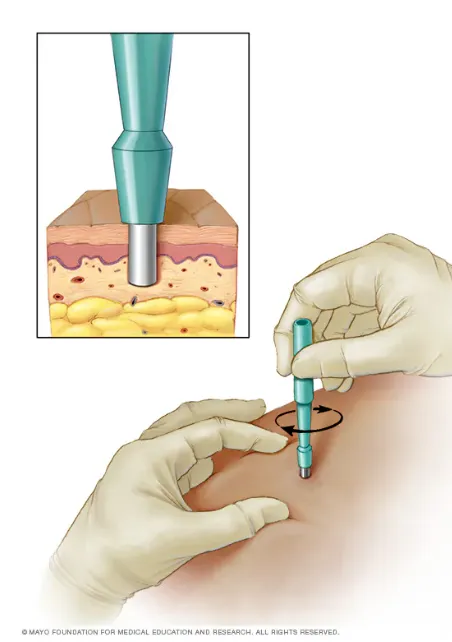
Fig. 18.1: Skin Punch Biopsy
Source: Mayo Clinic — Skin Biopsy
Post-procedure:
-
Send specimen in formalin (or Michel’s medium for immunofluorescence).
-
Wound care instructions and follow-up.
3. Cryotherapy and Electrocautery
Section titled “3. Cryotherapy and Electrocautery”Cryotherapy
Section titled “Cryotherapy”-
Mechanism: Rapid freezing with liquid nitrogen causes cellular destruction.
-
Indications:
-
Viral warts
-
Actinic keratoses
-
Seborrheic keratoses
-
-
Technique:
-
Apply liquid nitrogen via cotton-tipped applicator or cryospray.
-
Freeze-thaw cycles (usually 2–3).
-
-
Side effects:
- Pain, blistering, hypopigmentation, scarring.
Electrocautery
Section titled “Electrocautery”-
Mechanism: High-frequency electric current causes tissue coagulation or destruction.
-
Indications:
-
Skin tags
-
Small benign lesions
-
Haemostasis during minor procedures
-
-
Types:
-
Electrofulguration (superficial)
-
Electrodesiccation (deeper)
-
-
Precautions:
-
Avoid in patients with pacemakers.
-
Use a grounding pad if monopolar.
-
4. Patch Testing (Introduction)
Section titled “4. Patch Testing (Introduction)”Purpose:
- To identify delayed-type (Type IV) hypersensitivity reactions in allergic contact dermatitis.
Procedure:
-
Standardized allergens applied to the back using adhesive patches.
-
Patches remain in place for 48 hours.
-
Readings taken at 48 and 72–96 hours.
Interpretation:
-
Positive reaction: erythema, papules, vesicles at test site.
-
Graded as + (mild) to +++ (strong).
Common allergens:
- Nickel, fragrance mix, preservatives, rubber chemicals.
Limitations:
-
Cannot detect irritant reactions.
-
False positives/negatives possible.
Case-Based Discussions
Section titled “Case-Based Discussions”Case 1: Skin Scrapings and KOH Prep
Section titled “Case 1: Skin Scrapings and KOH Prep”Scenario: A 10-year-old boy presents with a scaly, itchy patch on his scalp with hair loss. You suspect tinea capitis.
Discussion Points:
-
What is the most appropriate diagnostic test? → Skin scraping from the active edge for KOH prep.
-
What findings would confirm your diagnosis? → Septate hyphae under microscopy.
-
What are the limitations of this test? → False negatives if scraping is inadequate or the sample is old.
Case Study: Punch Biopsy (Observation)
Section titled “Case Study: Punch Biopsy (Observation)”Scenario: A 55-year-old woman presents with a non-healing, pigmented lesion on her forearm. The dermatologist plans a punch biopsy.
Discussion Points:
-
Why is a punch biopsy preferred here? → Full-thickness sample for histopathology to rule out malignancy.
-
What are the key steps in the procedure? → Local anaesthesia → punch tool → sample removal → haemostasis → suture.
-
What are the risks? → Bleeding, infection, scarring.
Case Study: Cryotherapy
Section titled “Case Study: Cryotherapy”Scenario: A 30-year-old man presents with multiple plantar warts. The dermatologist recommends cryotherapy.
Discussion Points:
-
How does cryotherapy work? → Freezing causes cellular destruction and immune stimulation.
-
What are the expected side effects? → Pain, blistering, hypopigmentation.
-
When should it be avoided? → In patients with cold intolerance, Raynaud’s, or poor circulation.
Case Study: Patch Testing
Section titled “Case Study: Patch Testing”Scenario: A 40-year-old woman presents with chronic hand eczema. You suspect allergic contact dermatitis.
Discussion Points:
-
What is the role of patch testing? → Identifies delayed hypersensitivity to specific allergens.
-
How is it performed? → Allergens applied to back → left for 48 hours → readings at 48 and 72–96 hours.
-
What are common allergens? → Nickel, fragrance mix, preservatives, rubber chemicals.
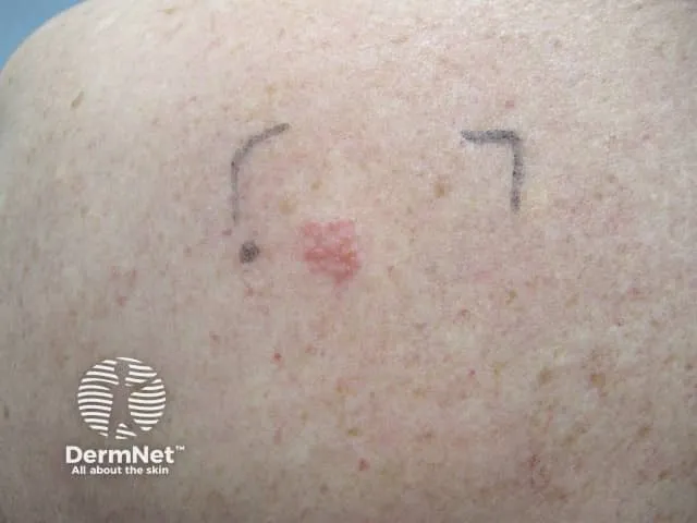
Fig. 18.2: Patch Test on the Back — Day 5 (contact dermatitis, nickel contact allergy)
Image sourced from DermNet.
Procedure Checklists
Section titled “Procedure Checklists”1. Skin Scrapings and KOH Prep
Section titled “1. Skin Scrapings and KOH Prep”Table 18.3: Skin Scrapings and KOH Prep — Step-by-Step
| Step | Description |
|---|---|
| 1 | Clean the lesion with an alcohol swab |
| 2 | Use a scalpel to scrape the active edge of the lesion |
| 3 | Collect scales on a glass slide |
| 4 | Add 10–20% KOH and cover with a coverslip |
| 5 | Gently heat or wait 15–30 minutes |
| 6 | Examine under a microscope for fungal elements |
2. Punch Biopsy
Section titled “2. Punch Biopsy”Table 18.4: Punch Biopsy — Step-by-Step
| Step | Description |
|---|---|
| 1 | Obtain informed consent |
| 2 | Clean the area and inject local anaesthetic |
| 3 | Use a punch tool (2–6 mm) to core the lesion |
| 4 | Lift the sample with forceps and cut the base |
| 5 | Achieve haemostasis (pressure or suture) |
| 6 | Place the specimen in formalin |
| 7 | Provide wound care instructions |
3. Cryotherapy
Section titled “3. Cryotherapy”Table 18.5: Cryotherapy — Step-by-Step
| Step | Description |
|---|---|
| 1 | Explain the procedure and obtain consent |
| 2 | Clean the lesion area |
| 3 | Apply liquid nitrogen using a cotton tip or cryospray |
| 4 | Freeze for 10–30 seconds (until halo forms) |
| 5 | Allow thawing; repeat if needed |
| 6 | Advise on post-procedure care (blistering, pain) |
4. Patch Testing
Section titled “4. Patch Testing”Table 18.6: Patch Testing — Step-by-Step
| Step | Description |
|---|---|
| 1 | Identify suspected allergens |
| 2 | Apply standardized allergens to the back using a patch panel |
| 3 | Leave patches in place for 48 hours |
| 4 | Remove and perform the first reading at 48 hours |
| 5 | Perform second reading at 72–96 hours |
| 6 | Grade reactions and interpret results |
| 7 | Counsel the patient on allergen avoidance |
Multiple Choice Questions
Section titled “Multiple Choice Questions”Tap an answer to check yourself — the correct option and an explanation appear once you choose.
Q1 A 12-year-old boy presents with a scaly, annular lesion on his arm. You suspect tinea corporis. What is the most appropriate diagnostic test?
KOH preparation is the first-line diagnostic test for superficial fungal infections. Scraping the active edge and applying KOH dissolves keratin and reveals fungal hyphae under microscopy. Skin biopsy is used for histopathological diagnosis of deeper or atypical lesions, Gram stain detects bacteria, and patch testing identifies contact allergens.
Q2 Which of the following is a correct indication for a punch biopsy?
A punch biopsy obtains a full-thickness skin sample for histopathological diagnosis and is indicated for suspected skin cancers such as basal cell carcinoma, inflammatory dermatoses, and bullous disorders. Superficial fungal infections are diagnosed with KOH prep, viral warts are treated with cryotherapy or electrocautery, and allergic contact dermatitis is diagnosed with patch testing.
Q3 Which of the following is a common side effect of cryotherapy?
Blistering is a well-recognised and common side effect of cryotherapy, occurring as a result of cellular destruction from rapid freezing with liquid nitrogen. Other expected side effects include pain, hypopigmentation, and scarring. Hyperpigmentation, alopecia, and keloid formation are not typical sequelae of cryotherapy.
Q4 Patch testing is most useful in diagnosing:
Patch testing detects delayed-type (Type IV) hypersensitivity reactions and is the investigation of choice for allergic contact dermatitis. It cannot distinguish irritant reactions (which are non-immunological), and is not the primary investigation for urticaria (which is IgE-mediated) or atopic dermatitis.
Short Answer Questions (SAQs)
Section titled “Short Answer Questions (SAQs)”SAQ 1: KOH Preparation
Section titled “SAQ 1: KOH Preparation”SAQ
Describe the steps involved in performing a KOH preparation for suspected dermatophytosis.
Reveal model answer
- Clean the lesion with alcohol.
- Scrape the active edge using a scalpel blade.
- Place the scales on a glass slide.
- Add 10–20% KOH solution and cover with a coverslip.
- Gently heat or wait 15–30 minutes.
- Examine under the microscope for long, branching septate hyphae (dermatophytes), budding yeast with pseudohyphae (Candida), or the “spaghetti and meatballs” pattern (Malassezia).
Pitfalls to avoid: scraping from the wrong site (scrape the active edge, not the centre) and misinterpreting keratin fibers as hyphae.
SAQ 2: Cryotherapy
Section titled “SAQ 2: Cryotherapy”SAQ
List three indications and three potential complications of cryotherapy.
Reveal model answer
Indications:
- Viral warts
- Actinic keratoses
- Seborrheic keratoses
Complications:
- Pain and blistering
- Hypopigmentation
- Scarring
Note: Cryotherapy should be avoided in patients with cold intolerance, Raynaud’s phenomenon, or poor peripheral circulation.
OSCE Stations
Section titled “OSCE Stations”OSCE 1: KOH Prep for Tinea Corporis
Section titled “OSCE 1: KOH Prep for Tinea Corporis”Scenario: A 9-year-old child presents with a ring-shaped, scaly lesion on the forearm. You suspect tinea corporis.
Task: Take a focused history. Demonstrate or describe how to perform a KOH prep. Explain the purpose of the test to the parent.
Self-assess against checklist
Tick each point you covered, then check your score.
- Asks about onset, progression, and exposure history (animals, contacts with similar rash).
- Identifies the lesion as annular and scaly with active edge.
- Explains that the most appropriate diagnostic test is a KOH preparation.
- Describes cleaning the lesion with alcohol before scraping.
- States that scraping should be taken from the active edge of the lesion.
- Describes collecting scales onto a glass slide and adding 10–20% KOH.
- States that the preparation is gently heated or left for 15–30 minutes before microscopy.
- Explains that microscopy looks for branching septate hyphae to confirm a fungal infection.
- Advises the parent that results confirm the diagnosis and guide antifungal treatment.
- Advises on treatment pending results and basic hygiene measures to prevent spread.
OSCE 2: Punch Biopsy Observation
Section titled “OSCE 2: Punch Biopsy Observation”Scenario: A 60-year-old man presents with a non-healing pigmented lesion on the cheek. The dermatologist plans a punch biopsy.
Task: Observe the procedure. Describe the steps and rationale. Explain post-procedure care to the patient.
Self-assess against checklist
Tick each point you covered, then check your score.
- States that a punch biopsy obtains a full-thickness skin sample for histopathological diagnosis.
- Explains that the indication is to rule out malignancy (e.g., melanoma, BCC, SCC).
- Describes obtaining informed consent before the procedure.
- Describes cleaning the area and injecting local anaesthetic (lignocaine).
- Explains that a circular punch tool (2–6 mm) is used to core out the lesion.
- Describes lifting the sample with forceps and cutting it at the base.
- States that haemostasis is achieved with pressure or a suture.
- Explains that the specimen is placed in formalin for histopathology (or Michel’s medium if immunofluorescence is required).
- Advises the patient on wound care and keeping the site clean and dry.
- Explains that follow-up is required to review histopathology results.
OSCE 3: Patch Testing Explanation
Section titled “OSCE 3: Patch Testing Explanation”Scenario: A 35-year-old woman with chronic hand eczema is referred for patch testing.
Task: Explain the purpose and process of patch testing. Describe how results are interpreted. Counsel on allergen avoidance.
Self-assess against checklist
Tick each point you covered, then check your score.
- Explains that patch testing identifies delayed-type (Type IV) hypersensitivity reactions in allergic contact dermatitis.
- Clarifies that patch testing is different from prick tests and does not detect irritant reactions.
- Describes that standardized allergens (e.g., nickel, fragrance mix, preservatives, rubber chemicals) are applied to the back using adhesive patches.
- States that patches remain in place for 48 hours.
- Explains that the first reading is taken at 48 hours when the patches are removed.
- States that a second reading is performed at 72–96 hours.
- Describes a positive reaction as erythema, papules, or vesicles at the test site, graded + to +++.
- Counsels the patient to avoid confirmed allergens in daily products, cosmetics, and occupational exposures.
- Emphasizes the importance of follow-up and documentation of results.
Reference Handout: Free Dermatology Image Resources
Section titled “Reference Handout: Free Dermatology Image Resources”The notes in this book have been kept brief intentionally. The following list of images of skin disorders can be accessed during your revision of the subject. Please read the terms and conditions for the use of the images.
DermNet NZ
Section titled “DermNet NZ”Description: Comprehensive A–Z image library of skin conditions, searchable by lesion type, body location, and skin tone.
Access Link: https://dermnetnz.org/images
Usage Guidelines: Free for non-commercial educational use. Must credit ‘DermNet’ and link back to the site.
SCIN Dataset (Google Research)
Section titled “SCIN Dataset (Google Research)”Description: 10,000+ images of skin, nail, and hair conditions across diverse skin tones and body sites.
Access Link: https://research.google/blog/scin-a-new-resource-for-representative-dermatology-images/
Usage Guidelines: Open-access for education and research. Includes metadata and dermatologist labels.
JDD Inclusive Derm Atlas
Section titled “JDD Inclusive Derm Atlas”Description: 1,000+ royalty-free images of 110+ conditions across all skin tones with expert commentary.
Access Link: https://www.jddonline.com/project-atlas
Usage Guidelines: Free for non-commercial educational use. Real patient images with diagnostic insights.
MedPix (U.S. National Library of Medicine)
Section titled “MedPix (U.S. National Library of Medicine)”Description: 59,000+ medical images and 12,000+ case scenarios across specialties, including dermatology.
Access Link: https://medpix.nlm.nih.gov/home
Usage Guidelines: Free for educational use. Includes case-based learning tools.
ISIC Archive
Section titled “ISIC Archive”Description: Open-source archive of skin lesion images, focused on dermoscopy and skin cancer diagnosis.
Access Link: https://www.isic-archive.com/
Usage Guidelines: Public resource for teaching, research, and AI development.
Further Reading
Section titled “Further Reading”-
Bolognia JL, Schaffer JV, Cerroni L. Dermatology. 5th ed. Elsevier; 2024.
-
Griffiths CEM, Barker J, Bleiker T, Chalmers R, Creamer D. Rook’s Textbook of Dermatology. 10th ed. Wiley-Blackwell; 2024.
-
Gniadecki R. A decade of progress and innovation in dermatology. Front Med (Lausanne). 2025 Jan 21;11:1546471. doi: 10.3389/fmed.2024.1546471.
-
McGrath LN. A year in review: new treatments and expanded indications in dermatology 2024. Dermatol Ther. 2025 Mar;35(2):e15465. doi: 10.1111/dth.15465.
-
Sebaratnam DF, et al. Rook’s Textbook of Dermatology, 10th edition: A focus on clinical guidelines and treatment algorithms. Br J Dermatol. 2024 May;191(5):854–862. doi: 10.1111/bjd.23753.