Module 10: Skin Manifestations of Systemic Diseases in Malaysia
Introduction
Section titled “Introduction”This module highlights additional systemic conditions where skin findings are often the first clue to underlying systemic diseases common in Malaysia, such as Henoch-Schönlein Purpura (HSP), Dengue Fever, Rheumatic Fever, and HIV/AIDS. Recognizing these cutaneous signs helps in early diagnosis and appropriate management.
Learning Objectives
Section titled “Learning Objectives”-
Identify characteristic skin manifestations of key systemic diseases.
-
Understand the pathophysiologic mechanisms linking skin and systemic involvement.
-
Recognize clinical presentations and perform relevant investigations.
-
Review management approaches for both cutaneous and systemic aspects.
-
Apply clinical knowledge through case-based learning.
1. Henoch-Schönlein Purpura (HSP)
Section titled “1. Henoch-Schönlein Purpura (HSP)”Pathophysiology
- IgA immune complex deposition causing small vessel vasculitis.
Clinical Features
-
Palpable purpura mainly on the lower limbs and buttocks.
-
Accompanied by arthralgia, abdominal pain, and renal involvement.
-
2–3 weeks of fever, headache, muscle/joint aches, or abdominal pain precede the symptoms.
The classic presentation of HSP is with a tetrad of symptoms and signs:
-
Rash
-
Arthritis
-
Abdominal pain
-
Kidney impairment
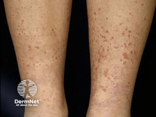
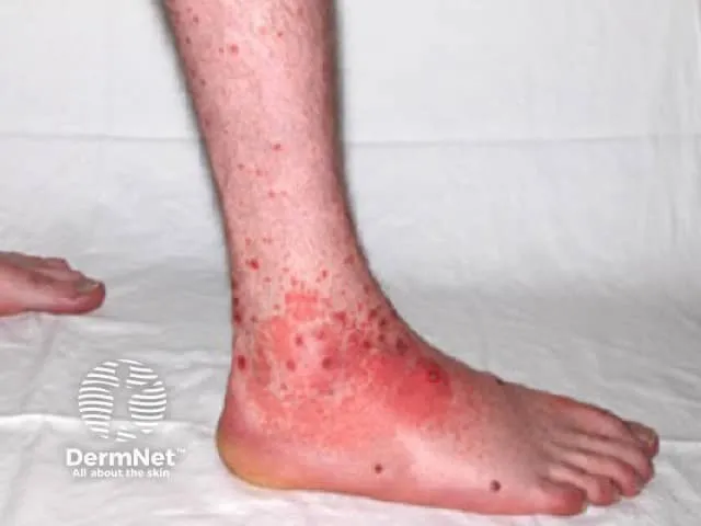
Fig. 10.1: Purpura on legs
Image sourced from DermNet.
Diagnosis
-
Clinical skin examination.
-
Skin or renal biopsy confirming leukocytoclastic vasculitis with IgA deposits.
-
Urinalysis for kidney involvement.
Treatment
-
Supportive care (rest, hydration).
-
NSAIDs for joint pain.
-
Corticosteroids if severe abdominal or renal symptoms.
2. Dengue Fever
Section titled “2. Dengue Fever”Pathophysiology
- Viral infection causing plasma leakage and thrombocytopenia.
Clinical Features
-
Maculopapular rash appearing on days 3–6.
-
Petechiae, purpura, and mucosal bleeding in severe cases.
-
Positive tourniquet test.
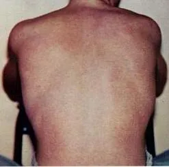
Fig. 10.2: Typical rash seen in dengue fever
Image sourced from Wikipedia.
Diagnosis
The Malaysian Clinical Practice Guidelines (CPG) for dengue diagnosis recommend:
-
Diagnostic tests include NS1 antigen test, PCR, or IgM serology to confirm dengue infection.
-
Frequent monitoring of platelet counts and haematocrit is essential to assess disease progression and risk of severe dengue.
-
Clinical evaluation involves history taking, physical exam, and full blood count (FBC), including platelet levels, with repeated FBC on day 3 and in patients with warning signs.
-
Diagnosis combines clinical features and laboratory confirmation for accurate disease phase assessment and timely management.
Treatment
-
Supportive care with fluids and monitoring.
-
Avoid NSAIDs due to bleeding risk.
3. Rheumatic Fever
Section titled “3. Rheumatic Fever”Pathophysiology
- Post-streptococcal autoimmune reaction causing inflammation in the heart, joints, CNS, and skin.
Clinical Features
-
Polyarthritis: inflammation of multiple joints (ankles, knees, elbows, wrists), can migrate between joints (migratory).
-
Carditis: inflammation of the heart valves, muscle, and surrounding membrane.
-
Sydenham chorea: nervous system disorder causing personality changes, muscle weakness, and involuntary movements.
-
Erythema marginatum: nonpruritic pink ring-shaped rash with central clearing.
-
Subcutaneous nodules on pressure areas.
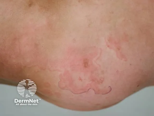
Fig. 10.3: Erythema marginatum
Image sourced from DermNet.
Diagnosis
Jones criteria (clinical features + evidence of recent streptococcal infection).
The modified Jones criteria require:
-
Major criteria: carditis, arthritis, chorea, erythema marginatum, subcutaneous nodules.
-
Minor criteria: polyarthralgia, fever ≥ 38.5°C, elevated inflammatory markers (ESR or CRP), prolonged PR interval on ECG.
-
For diagnosis, either 2 major criteria, or 1 major and 2 minor criteria must be met, along with evidence of preceding streptococcal infection confirmed by throat culture, rapid antigen test, or elevated antistreptococcal antibody titres (such as antistreptolysin O).
Treatment
-
Penicillin to eradicate streptococcus.
-
Aspirin/corticosteroids to control inflammation.
4. HIV/AIDS
Section titled “4. HIV/AIDS”Pathophysiology
- Immunosuppression leading to opportunistic infections and neoplasms.
Clinical Features
-
Kaposi sarcoma: violaceous plaques/nodules.
-
Seborrhoeic dermatitis, molluscum contagiosum, oral hairy leukoplakia.
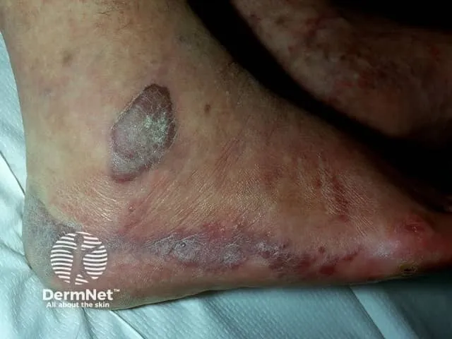
Fig. 10.4: Kaposi sarcoma
Image sourced from DermNet.
Diagnosis
-
HIV testing should be offered routinely to individuals with symptoms or conditions that might indicate HIV, in line with HIV indicator conditions.
-
HIV serology is the primary diagnostic tool comprising initial antigen-antibody combination tests (fourth/fifth generation) to detect both HIV antibodies and p24 antigen.
-
Confirmation of positive results should be done with supplementary tests such as immunoblot assays.
-
Testing is also recommended for people with neoplastic lesions such as Kaposi sarcoma due to HIV association, where skin biopsy may be done to confirm these lesions.
-
HIV testing should be offered in high-risk clinical settings (e.g., sexual health clinics, antenatal care, substance misuse services) and to patients with specific indicator conditions.
Treatment
-
Antiretroviral therapy (ART).
-
Treat skin conditions as per individual diagnosis.
Multiple Choice Questions
Section titled “Multiple Choice Questions”Tap an answer to check yourself — the correct option and an explanation appear once you choose.
Q1 What is the typical rash distribution in Henoch-Schönlein Purpura (HSP)?
Palpable purpura on the lower limbs and buttocks is classic for HSP, reflecting small vessel vasculitis caused by IgA immune complex deposition in dependent areas. Generalised maculopapular rash is more typical of dengue; vesicular trunk rash suggests varicella; target lesions on palms are seen in erythema multiforme.
Q2 When does the dengue maculopapular rash usually appear?
The maculopapular rash of dengue fever characteristically appears around days 3–6 of illness, coinciding with defervescence. Rash on day 1 is not typical; a rash appearing after day 10 or post-recovery would suggest an alternative diagnosis.
Q3 Which is the characteristic skin rash of acute rheumatic fever?
Erythema marginatum — a non-pruritic, pink ring-shaped rash with central clearing — is one of the five major Jones criteria for rheumatic fever. Kaposi sarcoma is associated with HIV, seborrhoeic dermatitis with Malassezia overgrowth, and livedo reticularis with vascular conditions such as antiphospholipid syndrome.
Q4 Which is a common cutaneous manifestation of HIV/AIDS?
Kaposi sarcoma — presenting as violaceous plaques or nodules — is a hallmark neoplasm of advanced HIV infection caused by human herpesvirus 8 (HHV-8) in the setting of immunosuppression. Subcutaneous nodules and palpable purpura are features of rheumatic fever and HSP respectively; petechiae are seen in dengue.
Q5 What is the mainstay treatment for acute rheumatic fever?
Penicillin is essential to eradicate the precipitating group A streptococcal infection. Anti-inflammatory agents (aspirin or corticosteroids) are added for carditis and arthritis, but penicillin remains the cornerstone because eradicating streptococcus is required to halt the autoimmune response. Antiretroviral therapy is for HIV; supportive hydration is the mainstay for dengue.
OSCE Stations
Section titled “OSCE Stations”OSCE 1: Henoch-Schönlein Purpura
Section titled “OSCE 1: Henoch-Schönlein Purpura”Scenario: A 12-year-old boy presents with palpable purpura predominantly on his legs, abdominal pain, and joint swelling. He had an upper respiratory tract infection 2 weeks ago.
Task: Take a focused history, examine the patient, establish the diagnosis, and outline your investigation and management plan.
Self-assess against checklist
Tick each point you covered, then check your score.
- Takes history of recent upper respiratory tract infection and timeline of symptoms.
- Asks about rash evolution — onset, distribution, and character (palpable vs. flat).
- Enquires about joint swelling and pain, abdominal pain, and urinary symptoms (haematuria, proteinuria).
- Examines skin for palpable purpura over lower limbs and buttocks.
- Assesses musculoskeletal system for arthritis.
- Identifies the HSP tetrad: rash, arthritis, abdominal pain, renal involvement.
- Explains IgA immune complex vasculitis as the underlying mechanism.
- Orders urinalysis to assess renal involvement; arranges skin or renal biopsy if indicated.
- Plans treatment: supportive rest and hydration, NSAIDs for arthralgia, corticosteroids if severe GI or renal symptoms.
- Counsels family on prognosis and the importance of monitoring kidney function.