Module 17: Leprosy (Hansen's Disease)
1. Learning Outcomes
Section titled “1. Learning Outcomes”By the end of this session, students should be able to:
-
Explain the pathogenesis, transmission, and classification of leprosy.
-
Identify clinical features and complications, and outline diagnostic approaches.
-
Discuss prevention strategies and current WHO-recommended treatment protocols.
2. Introduction & Prevalence
Section titled “2. Introduction & Prevalence”-
Leprosy is a chronic infectious disease caused by Mycobacterium leprae.
-
Affects skin, peripheral nerves, eyes, and upper respiratory mucosa.
-
Transmission: Prolonged close contact via nasal droplets from untreated patients.
-
Global burden: ~200,000 new cases annually; endemic in India, Brazil, Indonesia.
-
Malaysia: 66 new cases reported by May 2025; outbreaks in Negeri Sembilan and Sabah.
3. Aetiology
Section titled “3. Aetiology”-
Caused by Mycobacterium leprae, an acid-fast bacillus.
-
Incubation period: 5–20 years.
-
Cannot be cultured in vitro; diagnosis is clinical and histopathological.
-
Transmission requires prolonged exposure; it is not spread by casual contact.
4. Diagnosis
Section titled “4. Diagnosis”Clinical Features
-
Skin lesions: Hypopigmented or erythematous patches with sensory loss.
-
Peripheral nerve involvement: Thickened nerves, muscle weakness, paresthesia.
-
Ocular signs: Lagophthalmos, corneal ulcers, blindness.
-
Other signs: Madarosis, nasal collapse, claw hand.
Cardinal Signs (WHO)
-
Definite sensory loss in skin patch.
-
Thickened/enlarged peripheral nerve with sensory/motor loss.
-
Presence of bacilli in slit-skin smear.
Classification
-
Paucibacillary (PB): ≤5 lesions, no bacilli.
-
Multibacillary (MB): >5 lesions, bacilli present or nerve involvement.
Diagnosis confirmed by one of:
Section titled “Diagnosis confirmed by one of:”-
Skin Slit Smear: Dermis smear checked for acid-fast bacilli; for multibacillary leprosy.
-
Lepromin Test: Intradermal test to classify leprosy type.
-
Skin Biopsy: Shows leprosy features; may need special stains.
-
M. leprae DNA PCR: Highly specific for leprosy detection.
Images
Section titled “Images”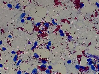
Fig. 17.1: M. leprae acid-fast bacilli (Ziehl–Neelsen stain)
As an acid-fast bacterium, M. leprae appears red when a Ziehl–Neelsen stain is used.
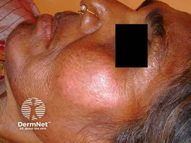
Fig. 17.2: Leprosy — Tuberculoid (well-defined erythematous plaque on the cheek)
Image sourced from DermNet.
Paucibacillary form is defined clinically by:
-
A few (1–2) sharply defined red patches with raised borders or a single larger hypopigmented patch less than 10 cm in diameter
-
Loss of sweating with rough, dry hairless skin in the patches
-
Loss of sensation in lesions
-
Affected nerves are thickened and tender on palpation
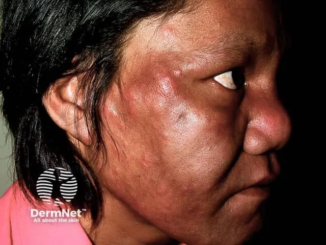
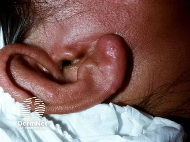
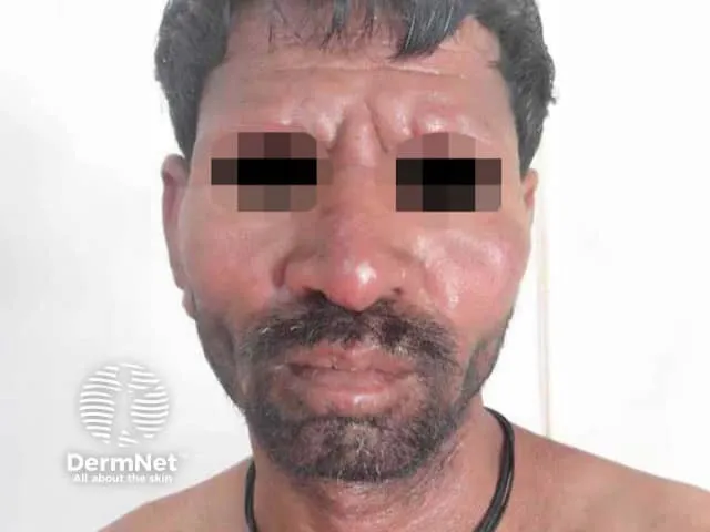
Fig. 17.3: Lepromatous leprosy (i), (ii), (iii)
Image sourced from DermNet.
Multibacillary form is defined by:
-
Early symptoms: nasal stuffiness, discharge, and bleeding
-
Swelling and thickening of limbs
-
Widespread poorly defined hypopigmented and erythematous macules; shiny surface, sensation may be normal
-
Progresses to infiltration of skin (nodules and plaques)
-
Characteristic leonine facies with thickening of the forehead, loss of eyebrows and eyelashes (madarosis), distortion of the nose, and thickening of the earlobes
-
Involvement of other systems
Table 17.1: Differential Diagnosis of Hypopigmented Patches and Macules
| Differential Diagnosis | Clinical Features | Key Distinguishing Points | Diagnostic Tests/Clues |
|---|---|---|---|
| Pityriasis alba | Hypopigmented, ill-defined scaly patches | Mostly in children, no sensory loss | Clinical appearance |
| Pityriasis versicolor | Hypo/hyperpigmented fine scaling patches | Caused by Malassezia yeast | KOH preparation showing yeast hyphae |
| Leishmaniasis | Ulcerative or plaque lesions | History of sandfly exposure | Skin biopsy, smear to identify parasites |
| Yaws | Papillomatous or ulcerative lesions in children | Tropical endemic area | Serology tests |
| Cutaneous leishmaniasis | Chronic ulcer/plaque, raised borders | Endemic region, raised lesion borders | Parasite demonstration in biopsy/smear |
| Lupus vulgaris (cutaneous tuberculosis) | Slowly progressive plaques, apple jelly nodules on diascopy | Positive tuberculin skin test | Skin biopsy showing caseating granulomas |
| Leprosy | Hypopigmented or erythematous patches, loss of sensation, thickened nerves | Peripheral nerve thickening, sensory/motor deficits, chronic onset | Acid-fast bacilli in skin smears/biopsy, clinical nerve exam |
| Fungal infections (e.g., tinea corporis) | Scaly plaques | No nerve involvement | KOH prep, fungal culture |
| Vitiligo | Depigmented well-demarcated patches | No sensory loss | Clinical appearance |
| Sarcoidosis | Papules or plaques | Non-caseating granulomas on biopsy | Skin biopsy histology |
5. Complications
Section titled “5. Complications”Neurological
Section titled “Neurological”-
Peripheral neuropathy → claw hand, foot drop.
-
Nerve abscesses.
Ocular
Section titled “Ocular”- Corneal ulcers, uveitis, blindness.
- Chronic ulcers, secondary infections.
Systemic
Section titled “Systemic”Lepra reactions:
-
Type 1 (Reversal): Inflammation of existing lesions, neuritis.
-
Type 2 (ENL): Painful nodules, fever, systemic symptoms.
Psychosocial
Section titled “Psychosocial”- Stigma, discrimination, depression.
6. Prevention & Treatment
Section titled “6. Prevention & Treatment”Prevention
Section titled “Prevention”-
Early detection and MDT.
-
Contact tracing and Single Dose Rifampicin (SDR-PEP) for contacts.
-
BCG vaccine offers partial protection.
-
Community education to reduce stigma.
Treatment (WHO MDT Guidelines 2025)
Section titled “Treatment (WHO MDT Guidelines 2025)”-
PB Leprosy:
-
Duration: 6 months.
-
Drugs: Rifampicin (monthly) + Dapsone (daily).
-
-
MB Leprosy:
-
Duration: 12 months.
-
Drugs: Rifampicin (monthly) + Dapsone (daily) + Clofazimine (daily + monthly).
-
-
MDT is free via WHO and MOH Malaysia.
-
Lepra reactions:
-
Type 1: Prednisolone.
-
Type 2: Thalidomide (if available), corticosteroids.
-
7. Expanded Treatment for Leprosy
Section titled “7. Expanded Treatment for Leprosy”MDT is the standard treatment for all forms of leprosy. It prevents drug resistance, ensures cure, and reduces transmission. It is supplied free of charge by WHO to endemic countries including Malaysia.
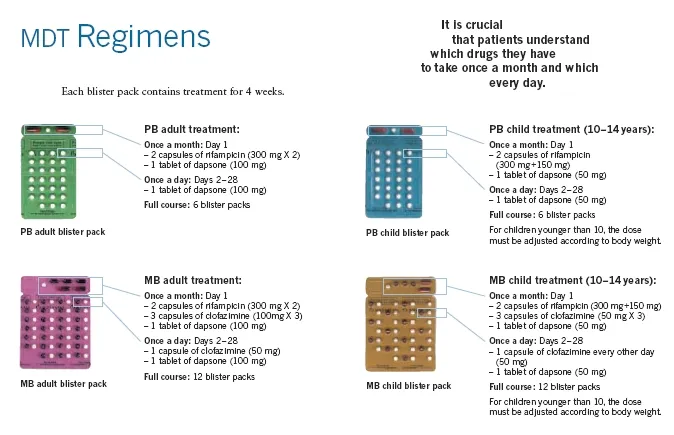
Fig. 17.4: WHO MDT blister pack for leprosy
Paucibacillary (PB) Leprosy
Section titled “Paucibacillary (PB) Leprosy”Definition: ≤5 skin lesions, no bacilli on slit-skin smear.
Duration: 6 months
Drug Regimen:
-
Rifampicin: 600 mg once monthly (supervised)
-
Dapsone: 100 mg daily (self-administered)
-
Clofazimine: 50 mg daily + 300 mg once monthly (added in 2025 update)
Notes:
-
Rifampicin may cause reddish urine temporarily.
-
Clofazimine may cause skin discoloration and dryness.
-
Dapsone is generally safe; monitor for sulfa allergy.
Multibacillary (MB) Leprosy
Section titled “Multibacillary (MB) Leprosy”Definition: >5 skin lesions, bacilli present, or nerve involvement.
Duration: 12 months
Drug Regimen:
-
Rifampicin: 600 mg once monthly (supervised)
-
Dapsone: 100 mg daily (self-administered)
-
Clofazimine: 50 mg daily + 300 mg once monthly (self-administered + supervised)
Notes:
-
MDT is highly effective in killing M. leprae.
-
Adherence is crucial to prevent relapse and resistance.
-
MDT is available at all MOH facilities in Malaysia.
Management of Lepra Reactions
Section titled “Management of Lepra Reactions”Table 17.2: Management of Lepra Reactions
| Reaction | Type | Symptoms | Treatment |
|---|---|---|---|
| Type 1 (Reversal Reaction) | Occurs in borderline forms | Inflammation of existing lesions, neuritis | Prednisolone 40–60 mg/day tapered over weeks; continue MDT |
| Type 2 (ENL — Erythema Nodosum Leprosum) | Occurs in MB cases | Painful nodules, fever, systemic symptoms | Thalidomide 100–300 mg/day (if available); Prednisolone or Clofazimine (anti-inflammatory dose); supportive care |
Monitoring and Follow-Up
Section titled “Monitoring and Follow-Up”-
Monthly supervised doses.
-
Monitor for:
-
Drug side effects (rash, GI upset, pigmentation).
-
Nerve function (sensory/motor).
-
Signs of lepra reactions.
-
-
Post-treatment disability care and rehabilitation.
Multiple Choice Questions
Section titled “Multiple Choice Questions”Tap an answer to check yourself — the correct option and an explanation appear once you choose.
Q1 A 35-year-old rubber tapper from Kuala Pilah presents with numb hypopigmented patches on his arms and a thickened ulnar nerve. Slit-skin smear is negative. What is the most likely classification?
Paucibacillary leprosy is defined by ≤5 skin lesions with a negative slit-skin smear. This patient has hypopigmented patches with sensory loss and a thickened nerve but no detectable bacilli, fitting PB criteria. Multibacillary requires >5 lesions or bacilli. Tuberculoid and indeterminate are histopathological sub-classifications not used in the WHO two-tier (PB/MB) treatment system.
Q2 A 42-year-old man on MDT for MB leprosy develops painful nodules, fever, and joint pain. What is the most likely diagnosis?
Erythema nodosum leprosum (Type 2 reaction) occurs in multibacillary leprosy and presents with painful subcutaneous nodules, fever, and systemic features including joint pain. Type 1 (reversal reaction) causes inflammation of existing lesions and neuritis without the painful nodules. Drug allergy and secondary bacterial infection are less consistent with this specific constellation of findings during MDT.
Q3 Which of the following drugs used in MDT causes skin discoloration?
Clofazimine causes reddish-brown to black skin discoloration, which is a well-recognized and reversible side effect. Rifampicin may cause reddish urine (not skin). Dapsone is generally safe without pigmentary changes. Isoniazid is not part of the leprosy MDT regimen.
Short Answer Questions (SAQs)
Section titled “Short Answer Questions (SAQs)”SAQ 1: Cardinal Signs and Treatment of Paucibacillary Leprosy
Section titled “SAQ 1: Cardinal Signs and Treatment of Paucibacillary Leprosy”SAQ
A 28-year-old woman presents with a single hypopigmented patch on her thigh with sensory loss. There is no nerve thickening.
a) What are the cardinal signs of leprosy (WHO)?
b) What is the recommended treatment for this patient?
Reveal model answer
a) Cardinal signs of leprosy (WHO):
- Definite sensory loss in a skin patch.
- Thickened or enlarged peripheral nerve with associated sensory or motor loss.
- Presence of acid-fast bacilli in slit-skin smear.
One cardinal sign is sufficient to diagnose leprosy.
b) Treatment:
This patient has a single hypopigmented patch with sensory loss and a negative nerve examination — consistent with Paucibacillary (PB) leprosy (≤5 lesions, no bacilli).
Recommended regimen:
- Rifampicin 600 mg once monthly (supervised)
- Dapsone 100 mg daily (self-administered)
- Duration: 6 months
MDT is available free of charge at all MOH facilities in Malaysia. Counsel the patient on medication side effects (reddish urine from Rifampicin) and the importance of adherence to prevent relapse.
SAQ 2: Complications and Management of Advanced Leprosy
Section titled “SAQ 2: Complications and Management of Advanced Leprosy”SAQ
A 50-year-old man with multibacillary leprosy develops claw hand and foot drop.
a) What complications of leprosy have occurred in this patient?
b) How would you manage this patient?
Reveal model answer
a) Complications:
- Peripheral neuropathy — damage to ulnar and common peroneal nerves leads to claw hand and foot drop respectively.
- Deformity and disability — progressive without treatment.
- This patient is also at risk of ocular complications (lagophthalmos, corneal ulcers), chronic skin ulcers, and secondary infections.
- Psychosocial complications: stigma, depression, and social discrimination are significant burdens in leprosy.
b) Management:
- Continue MDT — MB regimen (Rifampicin + Dapsone + Clofazimine for 12 months); MDT kills M. leprae and prevents further nerve damage.
- Monitor for lepra reactions — particularly Type 1 (reversal) which causes additional neuritis; treat with Prednisolone if present.
- Nerve function assessment — regular sensory and motor testing to detect new nerve damage early.
- Physiotherapy — to maintain joint mobility and prevent further contracture.
- Orthotic support — splints and footwear adaptations for foot drop and claw hand.
- Surgical referral — if reconstructive or tendon-transfer surgery is appropriate.
- Post-treatment disability care and rehabilitation — coordinate with physiotherapy and occupational therapy.
- Psychosocial support — address stigma, counsel patient and family, and facilitate community re-integration.
OSCE Stations
Section titled “OSCE Stations”OSCE 1: Evaluation and Counselling of Suspected Leprosy
Section titled “OSCE 1: Evaluation and Counselling of Suspected Leprosy”Scenario: A 40-year-old plantation worker presents with numb skin patches and thickened nerves on examination.
Task: Take a focused history, examine the patient, and counsel on diagnosis, treatment, stigma, and contact tracing.
Self-assess against checklist
Tick each point you covered, then check your score.
- Asks about onset and duration of skin patches and numbness.
- Enquires about history of close contact with a known leprosy case.
- Asks about other symptoms: nasal discharge, eye changes, limb weakness.
- Examines skin lesions: number, distribution, hypopigmentation or erythema.
- Tests sensation in skin patches (light touch, pin-prick).
- Palpates peripheral nerves for thickening and tenderness (e.g., ulnar, common peroneal).
- Assesses for motor deficits: claw hand, foot drop.
- Explains the diagnosis clearly and addresses stigma: leprosy is curable with MDT.
- Outlines the MDT regimen appropriate to classification (PB 6 months or MB 12 months).
- Counsels on medication side effects (reddish urine from Rifampicin, skin darkening from Clofazimine).
- Advises that MDT is free at all MOH facilities.
- Discusses contact screening and SDR-PEP for close contacts.
- Emphasises adherence to prevent relapse and resistance.
- Provides psychosocial reassurance and addresses community stigma.