Module 12: Skin Cancer
Learning Outcomes
Section titled “Learning Outcomes”By the end of this module, students should be able to:
-
Describe the epidemiology and risk factors for BCC, SCC, and melanoma.
-
Explain the pathophysiology and histological features of common skin cancers.
-
Recognize clinical presentations and dermoscopic features of skin malignancies.
-
Outline diagnostic approaches, including biopsy and staging systems.
-
Discuss treatment options, including surgical and non-surgical modalities.
-
Educate patients on skin cancer prevention and follow-up care.
Pathophysiology
Section titled “Pathophysiology”Basal Cell Carcinoma (BCC)
Section titled “Basal Cell Carcinoma (BCC)”-
Originates from basal keratinocytes of the epidermis, hair follicles, and sweat ducts.
-
UVB radiation causes mutations in PTCH1 and p53 genes.
-
Requires stromal support, hence rarely metastasizes (<1%).
-
Histologic subtypes:
-
Nodular: Pearly papule with rolled borders and telangiectasia.
-
Superficial: Scaly plaque with threadlike borders.
-
Pigmented: Melanin-rich, mimics melanoma.
-
Morpheaform: Indistinct margins, aggressive.
-
Squamous Cell Carcinoma (SCC)
Section titled “Squamous Cell Carcinoma (SCC)”-
Arises from epidermal keratinocytes.
-
Progresses from actinic keratosis or Bowen’s disease.
-
UV radiation, chronic inflammation, immunosuppression, and HPV are key triggers.
-
Histologic subtypes:
-
Well-differentiated: Keratin pearls.
-
Spindle cell, Adenoid, Verrucous: Variable aggressiveness.
-
-
Metastasis risk: ~2–5%, higher in immunosuppressed patients.
Melanoma
Section titled “Melanoma”-
Malignant tumor of melanocytes.
-
UV exposure, genetic mutations (e.g., BRAF, CDKN2A), and fair skin are major risk factors.
-
Histologic types:
-
Superficial spreading: Radial growth, good prognosis.
-
Nodular: Vertical growth, poor prognosis.
-
Acral lentiginous: Palms, soles, nail beds; common in darker skin.
-
Lentigo maligna: Elderly, sun-exposed areas.
-
Diagnosis
Section titled “Diagnosis”Table 12.1: Clinical Features
| Type | Appearance | Common Sites |
|---|---|---|
| BCC | Pearly papule, rolled edges, telangiectasia | Face, neck |
| SCC | Crusted, ulcerated lesion, firm nodule | Sun-exposed areas |
| Melanoma | Irregular pigmented lesion, ABCDE criteria | Anywhere, including mucosa |
ABCDE Criteria for Melanoma
-
Asymmetry
-
Border irregularity
-
Colour variation
-
Diameter >6 mm
-
Evolution (change over time)
Investigations
Section titled “Investigations”-
Dermoscopy
-
Skin biopsy: Shave or punch depending on lesion type
-
Histopathology
-
Staging:
-
Clark’s levels: Depth of invasion
-
Breslow thickness: Prognostic indicator
-
TNM classification
-
Treatment
Section titled “Treatment”Basal Cell Carcinoma
Section titled “Basal Cell Carcinoma”-
Surgical excision: First line
-
Mohs micrographic surgery: For high-risk or facial lesions
-
Topical therapy: Imiquimod, 5-FU (for superficial BCC)
-
Cryotherapy, Photodynamic therapy, CO₂ laser: For selected cases
Squamous Cell Carcinoma
Section titled “Squamous Cell Carcinoma”-
Excision with clear margins
-
Mohs surgery: For aggressive or recurrent lesions
-
Radiotherapy: For inoperable cases
-
Topical agents: For SCC in situ (e.g., 5-FU, imiquimod)
Melanoma
Section titled “Melanoma”-
Wide local excision: Margin depends on Breslow depth
-
Sentinel lymph node biopsy: For staging
-
Immunotherapy: Checkpoint inhibitors (e.g., nivolumab)
-
Targeted therapy: BRAF/MEK inhibitors
-
Radiotherapy/Chemotherapy: For advanced disease
Prevention & Follow-Up
Section titled “Prevention & Follow-Up”-
Sun protection: SPF ≥30, protective clothing
-
Avoid tanning beds
-
Regular skin checks: Especially for high-risk individuals
-
Follow-up schedule:
-
Every 3–6 months for first 2 years
-
Annually thereafter
-
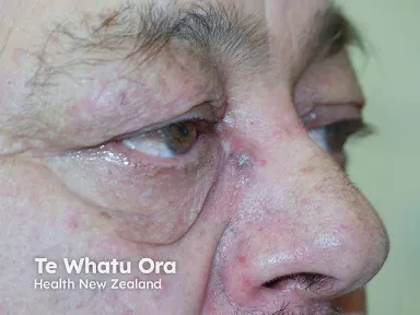
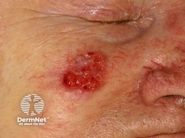
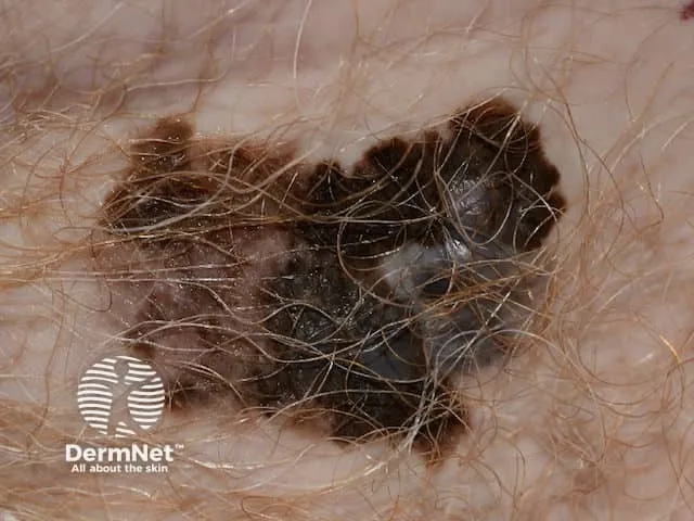
Fig. 12.1: Three skin cancers — pigmented basal cell carcinoma, squamous cell carcinoma, and melanoma
Image sourced from DermNet.
Multiple Choice Questions
Section titled “Multiple Choice Questions”Tap an answer to check yourself — the correct option and an explanation appear once you choose.
Q1 Which of the following is the most common skin cancer?
Basal cell carcinoma (BCC) is by far the most common skin cancer, accounting for approximately 70–80% of all skin malignancies. It arises from basal keratinocytes and is strongly associated with cumulative UV exposure, particularly on the face and neck.
Q2 Which histological feature is characteristic of squamous cell carcinoma?
Keratin pearls — concentric whorls of keratinizing squamous cells — are the hallmark of well-differentiated squamous cell carcinoma on histology. Palisading nuclei are seen in BCC, melanin granules in melanocytic lesions, and atypical melanocytes in melanoma.
Q3 Which of the following is a poor prognostic factor in melanoma?
Ulceration of a melanoma is an independent poor prognostic indicator and upstages the T category in the TNM system. Superficial spreading type carries a relatively good prognosis due to radial (rather than vertical) growth, and Breslow thickness <1 mm and Clark level II both indicate thin, early-stage disease with favourable outcomes.
Q4 Mohs micrographic surgery is most appropriate for which of the following?
Mohs surgery is indicated for BCC (and SCC) in high-risk, cosmetically sensitive, or functionally critical sites such as the nasal ala, eyelids, lips, and ears — where complete margin control and maximal tissue conservation are essential. Superficial BCC on the trunk can be managed with topical therapy or standard excision; melanoma requires wide local excision, not Mohs.
Q5 Which of the following is a known risk factor for squamous cell carcinoma?
Chronic wounds (e.g., Marjolin's ulcer), immunosuppression (e.g., transplant recipients), chronic UV exposure, HPV infection, and pre-existing lesions such as actinic keratosis and Bowen's disease are established risk factors for SCC. BRAF mutations are a driver of melanoma, not SCC, and topical corticosteroids are not a recognised cause.
Short Answer Questions
Section titled “Short Answer Questions”SAQ 1: ABCDE Criteria for Melanoma Screening
Section titled “SAQ 1: ABCDE Criteria for Melanoma Screening”SAQ
Describe the ABCDE criteria used in melanoma screening and explain the clinical significance of each parameter.
Reveal model answer
-
A — Asymmetry: One half of the lesion does not mirror the other; benign naevi are typically symmetrical.
-
B — Border: Irregular, scalloped, or poorly defined edges; melanomas lack the smooth, sharp border of benign lesions.
-
C — Colour: Variation in shades (brown, black, red, white, blue) within a single lesion; benign naevi are uniform in colour.
-
D — Diameter: Lesions greater than 6 mm warrant concern, though melanomas can be smaller at early stages.
-
E — Evolution: Any change in size, shape, colour, or new symptoms (bleeding, itching) over time is a red flag requiring urgent assessment.
SAQ 2: Histological Subtypes of Basal Cell Carcinoma
Section titled “SAQ 2: Histological Subtypes of Basal Cell Carcinoma”SAQ
List three histological subtypes of basal cell carcinoma and describe their clinical relevance.
Reveal model answer
-
Nodular BCC: Most common subtype; presents as a pearly papule with rolled borders and surface telangiectasia; typically well-circumscribed and amenable to standard excision.
-
Superficial BCC: Appears as a scaly, erythematous plaque with threadlike borders, often on the trunk; may be treated with topical imiquimod, 5-FU, or photodynamic therapy.
-
Morpheaform (sclerosing) BCC: Aggressive subtype with indistinct clinical margins and infiltrative growth; highest recurrence risk — Mohs micrographic surgery is the preferred treatment.
SAQ 3: Melanoma Staging
Section titled “SAQ 3: Melanoma Staging”SAQ
Outline the staging system used for melanoma, including the key parameters assessed.
Reveal model answer
-
Breslow thickness: Depth of tumour invasion measured in millimetres from the granular layer to the deepest tumour cell; the single most important prognostic factor.
-
Clark’s level: Anatomical level of invasion (I–V), from intraepidermal to subcutaneous fat; largely superseded by Breslow thickness but still reported.
-
TNM staging: Integrates primary tumour thickness and ulceration (T), regional lymph node involvement (N), and distant metastasis (M) to assign overall stage (0–IV).
-
Sentinel lymph node biopsy (SLNB): Performed for staging to detect lymph node spread and guide further management.
SAQ 4: Treatment of Squamous Cell Carcinoma
Section titled “SAQ 4: Treatment of Squamous Cell Carcinoma”SAQ
What are the treatment options for squamous cell carcinoma? Indicate when each is appropriate.
Reveal model answer
-
Surgical excision with clear margins: First-line for most SCC; margin width guided by tumour size and risk.
-
Mohs micrographic surgery: For high-risk, recurrent, or cosmetically/functionally critical site lesions (e.g., face, ears, hands).
-
Radiotherapy: For inoperable cases, elderly patients unsuitable for surgery, or as adjuvant therapy after excision with positive margins.
-
Topical agents (5-FU, imiquimod): Reserved for SCC in situ (Bowen’s disease) — not suitable for invasive SCC.
-
Systemic therapy: For locally advanced or metastatic disease not amenable to surgery or radiotherapy.
OSCE Stations
Section titled “OSCE Stations”OSCE 1: Basal Cell Carcinoma
Section titled “OSCE 1: Basal Cell Carcinoma”Scenario: A 65-year-old man presents with a pearly nodule on his right cheek that has slowly enlarged over 6 months.
Task: Take a focused history, perform a skin examination, suggest investigations, and outline management.
Self-assess against checklist
Tick each point you covered, then check your score.
- Asks about duration and rate of growth of the lesion.
- Enquires about sun exposure history, outdoor occupation, and use of sun protection.
- Asks about previous skin cancers, skin cancer in family members, and immunosuppression.
- Examines the lesion: identifies pearly surface, rolled (everted) borders, and surface telangiectasia.
- Checks for satellite lesions and regional lymph nodes.
- Requests dermoscopy to assess vascular pattern and pigmentation.
- Recommends punch or shave biopsy for histological confirmation.
- Discusses surgical excision as first-line treatment.
- Mentions Mohs micrographic surgery as preferred option for a facial high-risk lesion.
- Explains the importance of sun protection and regular follow-up for surveillance.
OSCE 2: Squamous Cell Carcinoma
Section titled “OSCE 2: Squamous Cell Carcinoma”Scenario: A 70-year-old woman with a history of renal transplant presents with a crusted lesion on her forearm.
Task: Assess risk factors, describe lesion characteristics, suggest diagnostic steps, and propose treatment.
Self-assess against checklist
Tick each point you covered, then check your score.
- Identifies immunosuppression (renal transplant) as a major risk factor for aggressive SCC.
- Asks about chronic sun exposure, previous actinic keratoses, and prior skin cancers.
- Asks about duration, growth rate, bleeding, and pain of the current lesion.
- Examines the lesion: describes crusted, ulcerated plaque with surrounding induration.
- Checks regional lymph nodes for metastatic spread.
- Recommends incisional or punch biopsy for histological diagnosis.
- Proposes surgical excision with adequate margins as primary treatment.
- Discusses adjuvant radiotherapy if margins are positive or the lesion is inoperable.
- Advises on the increased risk of multiple SCCs in immunosuppressed patients and the need for regular surveillance.
OSCE 3: Melanoma
Section titled “OSCE 3: Melanoma”Scenario: A 45-year-old woman presents with a pigmented lesion on her back that has changed in size and colour over 3 months.
Task: Apply ABCDE criteria, suggest investigations, discuss staging, and outline management.
Self-assess against checklist
Tick each point you covered, then check your score.
- Applies ABCDE criteria: notes asymmetry, irregular border, colour variation, diameter >6 mm, and recent evolution.
- Asks about personal or family history of melanoma and number of atypical naevi.
- Asks about sun exposure history and history of sunburn or tanning bed use.
- Performs full skin examination to identify any other suspicious lesions.
- Requests dermoscopy to assess dermoscopic features before biopsy.
- Recommends excisional biopsy with 1–2 mm margins for histological diagnosis and Breslow measurement.
- Explains that staging involves Breslow thickness, Clark level, TNM classification, and sentinel lymph node biopsy if indicated.
- Discusses wide local excision as the definitive surgical treatment with margins determined by Breslow depth.
- Mentions immunotherapy (checkpoint inhibitors) and targeted therapy (BRAF/MEK inhibitors) for advanced disease.
- Advises on follow-up schedule: every 3–6 months for the first 2 years, then annually.