Module 1: Dermatological Principles
1.1 Taking a Focused Dermatological History
Section titled “1.1 Taking a Focused Dermatological History”A comprehensive dermatological history is the cornerstone of diagnosis. It should be systematic, patient-centred, and clinically oriented.
Key Components
Section titled “Key Components”-
Chief Complaint
- Nature of the skin problem (e.g., rash, lesion, itch, discoloration).
-
History of Present Illness
-
Onset: Sudden or gradual?
-
Duration: Acute (<6 weeks) or chronic (>6 weeks)?
-
Progression: Static, spreading, or resolving?
-
Symptoms: Pruritus, pain, burning, bleeding, discharge.
-
Pattern: Intermittent or persistent?
-
Aggravating/Relieving Factors: Heat, cold, stress, sunlight, medications.
-
-
Past Dermatological History
- Previous similar episodes, treatments used, and response.
-
Medical and Surgical History
- Atopy, autoimmune diseases, diabetes, HIV, malignancies.
-
Drug History
- Prescription, OTC, herbal, recent changes.
-
Family History
-
Psoriasis, eczema, vitiligo, skin cancers.
Social and Occupational History- Exposure to chemicals, allergens, or infectious agents.
-
Travel History
- Especially relevant for tropical or parasitic skin diseases.
1.2 Structured Skin Examination Techniques
Section titled “1.2 Structured Skin Examination Techniques”A head-to-toe examination is essential. Use natural light or a dermatoscope as needed.
1. General Inspection
◦ Examine the entire skin surface, including the scalp, nails, mucosa, and intertriginous areas.
2. Lesion Assessment – “SALTSS” Mnemonic
◦ Site: Note the anatomical location and distribution.
◦ Arrangement: Identify if lesions are linear, grouped, annular, or dermatomal.
◦ Lesion Type: Determine if lesions are primary or secondary.
◦ Texture: Assess whether the surface is smooth, rough, or scaly.
◦ Size and Shape: Measure and describe.
◦ Surroundings: Look for erythema, oedema, or satellite lesions.
3. Palpation
◦ Feel for consistency, tenderness, temperature, mobility, and blanching.
4. Special Tests
◦ Diascopy: Apply a glass slide to assess blanching.
◦ Wood’s Lamp: Use for pigmentary disorders and infections.
◦ Dermatoscopy: Examine pigmented lesions and skin cancers.
1.3 Dermatological Terminology and Descriptions
Section titled “1.3 Dermatological Terminology and Descriptions”Precise terminology improves communication and documentation.
Table 1.1: Primary Lesions:
| Lesion | Description | Example |
|---|---|---|
| Macule | Flat, <1 cm | Freckle |
| Patch | Flat, >1 cm | Vitiligo |
| Papule | Raised, <1 cm | Wart |
| Plaque | Raised, >1 cm | Psoriasis |
| Vesicle | Fluid-filled, <1 cm | Herpes simplex |
| Bulla | Fluid-filled, >1 cm | Bullous pemphigoid |
| Pustule | Pus-filled | Acne |
| Nodule | Solid, deep | Dermatofibroma |
Secondary Lesions
Section titled “Secondary Lesions”-
Scale: Flakes of keratin (e.g., psoriasis)
-
Crust: Dried serum or pus (e.g., impetigo)
-
Erosion: Loss of epidermis
-
Ulcer: Loss of epidermis and dermis
-
Fissure: Linear crack
-
Scar: Fibrous tissue replacing normal skin
-
Lichenification: Thickened skin from chronic scratching
Distribution Patterns
Section titled “Distribution Patterns”-
Symmetrical: Psoriasis
[Update 2025 CDC/BASHH]: IL-17 and IL-23 inhibitors are preferred for biologic-naive patients with moderate-to-severe disease. -
Flexural: Atopic dermatitis
-
Extensor: Psoriasis
[Update 2025 CDC/BASHH]: IL-17 and IL-23 inhibitors are preferred for biologic-naive patients with moderate-to-severe disease. -
Dermatomal: Herpes zoster
-
Photo-distributed: Lupus erythematosus
Anatomy of the Skin and Clinical Correlation
Section titled “Anatomy of the Skin and Clinical Correlation”Understanding skin anatomy is crucial for localizing pathology and interpreting clinical signs (Fig. 1.1)
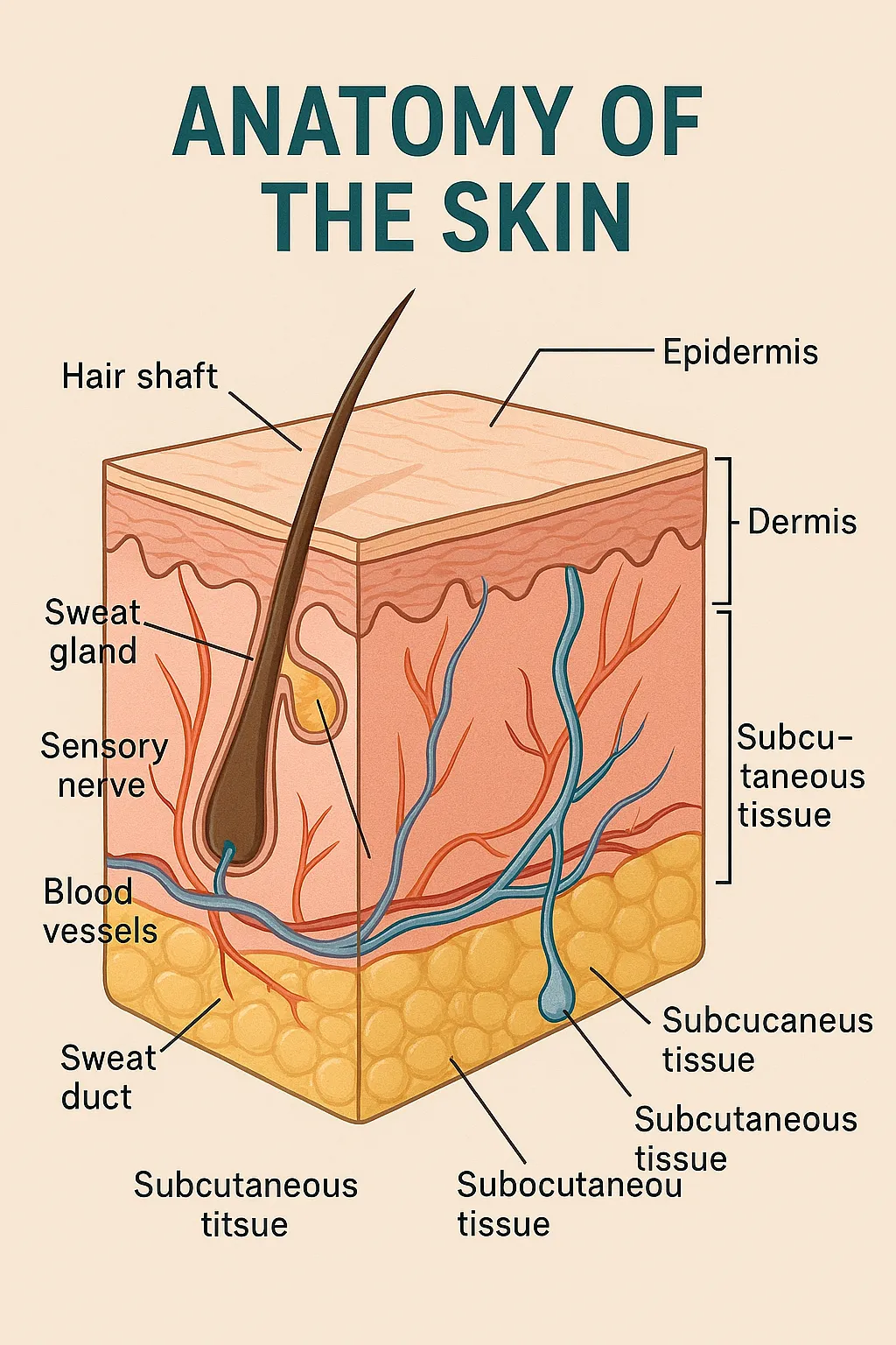
Fig. 1.1: Anatomy of the skin
1. Epidermis
-
Layers: Stratum corneum → lucidum → granulosum → spinosum → basale
-
Cells: Keratinocytes, melanocytes, Langerhans cells
-
Clinical relevance:
-
Psoriasis: Hyperproliferation of keratinocytes
-
Vitiligo: Loss of melanocytes
-
Eczema: Barrier dysfunction
-
2. Dermis
-
Components: Collagen, elastin, blood vessels, lymphatics, nerves
-
Structures: Hair follicles, sebaceous and sweat glands
-
Clinical relevance:
-
Urticaria: Vascular dilation
-
Lupus: Immune complex deposition
-
Cellulitis: Bacterial infection of dermis
-
3. Subcutaneous Tissue (Hypodermis)
-
Function: Insulation, energy storage, shock absorption
-
Clinical relevance:
-
Erythema nodosum: Inflammatory panniculitis
-
Lipomas: Benign fat tumours
-