Module 4: Skin Infections
Learning Objectives
Section titled “Learning Objectives”By the end of this module, students should be able to:
-
Describe the pathophysiology of common bacterial, viral, fungal, and parasitic skin infections.
-
Identify key clinical features and differentiate between types of skin infections.
-
Apply appropriate diagnostic methods for each condition.
-
Formulate evidence-based management plans, including pharmacological and non-pharmacological strategies.
-
Recognize complications and indications for referral or escalation of care.
-
Educate patients on prevention, hygiene, and treatment adherence.
Bacterial Infections
Section titled “Bacterial Infections”1. Impetigo
Section titled “1. Impetigo”-
Pathophysiology:
-
Superficial epidermal infection.
-
Caused by Staphylococcus aureus (bullous) or Streptococcus pyogenes (non-bullous).
-
Spread via direct contact or fomites.
-
-
Diagnosis:
-
Clinical: honey-coloured crusts, especially around the nose and mouth.
-
Swab for culture if resistant or recurrent.
-
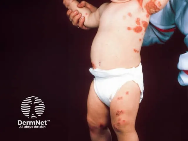
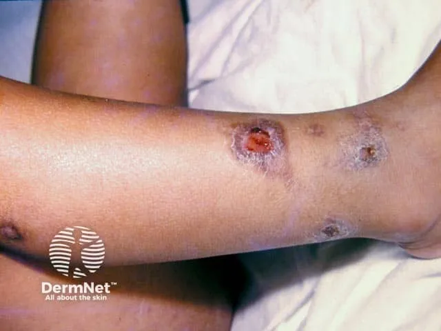
Fig. 4.1: Impetigo
- “Kissing lesions” on both sides of the axilla in impetigo
- Impetigo on the leg
Image sourced from DermNet.
-
Management:
-
Topical mupirocin or fusidic acid (mild).
-
Oral antibiotics (e.g., cloxacillin, cephalexin) for extensive or systemic involvement.
-
Hygiene education; avoid school/daycare until non-infectious.
-
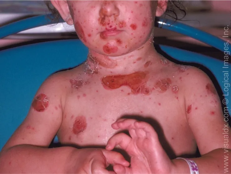
Fig. 4.2: Impetigo
2. Cellulitis
Section titled “2. Cellulitis”-
Pathophysiology:
-
Infection of the dermis and subcutaneous tissue.
-
Common organisms: Streptococcus pyogenes, S. aureus.
-
Entry via a skin breach (e.g., trauma, tinea pedis).
-
-
Diagnosis:
-
Clinical: erythema, warmth, swelling, tenderness, fever.
-
Blood cultures if febrile or immunocompromised.
-
-
Management:
-
Oral antibiotics (e.g., amoxicillin-clavulanate).
-
IV antibiotics for severe/systemic cases.
-
Elevation, rest, and treat the underlying cause (e.g., tinea).
-
Viral Infections
Section titled “Viral Infections”3. Herpes Simplex (HSV-1/2)
Section titled “3. Herpes Simplex (HSV-1/2)”-
Pathophysiology:
-
Primary infection followed by latency in sensory ganglia.
-
Reactivation triggered by stress, illness, UV light.
-
-
Diagnosis:
-
Clinical: grouped vesicles on an erythematous base, painful.
-
Tzanck smear (multinucleated giant cells); PCR for confirmation.
-
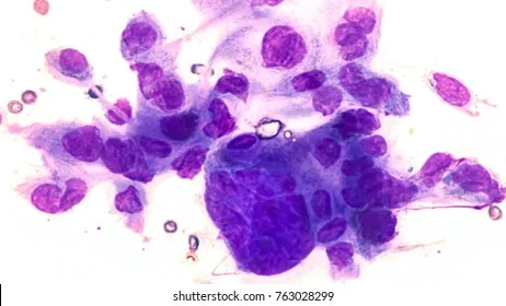
Fig. 4.3: Multinucleated giant cell (Tzanck smear)
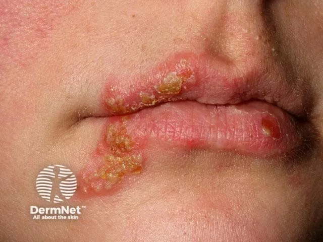
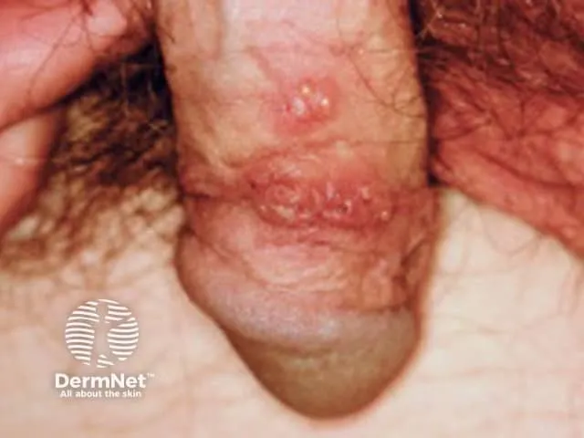
Fig. 4.4: (i) Herpes labialis and (ii) herpes genitalis
Image sourced from DermNet.
-
Management:
-
Oral acyclovir, valacyclovir (early initiation reduces duration).
-
Topical antivirals for mild cases.
-
Education on recurrence and transmission.
-
4. Varicella (Chickenpox)
Section titled “4. Varicella (Chickenpox)”-
Pathophysiology:
-
Primary infection with varicella-zoster virus (VZV).
-
Spread via respiratory droplets or direct contact.
-
-
Diagnosis:
-
Clinical: crops of vesicles at different stages, pruritic.
-
PCR or serology if atypical.
-
-
Management:
-
Supportive: antihistamines, calamine lotion, paracetamol.
-
Acyclovir for high-risk groups (e.g., immunocompromised, adults).
-
Isolation until lesions have crusted.
-
Fungal Infections
Section titled “Fungal Infections”5. Tinea (Dermatophytosis)
Section titled “5. Tinea (Dermatophytosis)”-
Pathophysiology:
-
Infection of keratinized tissue by dermatophytes (Trichophyton, Microsporum).
-
Spread via direct contact, fomites, animals.
-
-
Diagnosis:
-
KOH prep: hyphae visible.
-
Fungal culture for confirmation.
-
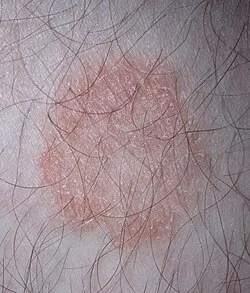
Fig. 4.5: Tinea (ringworm on a human leg)
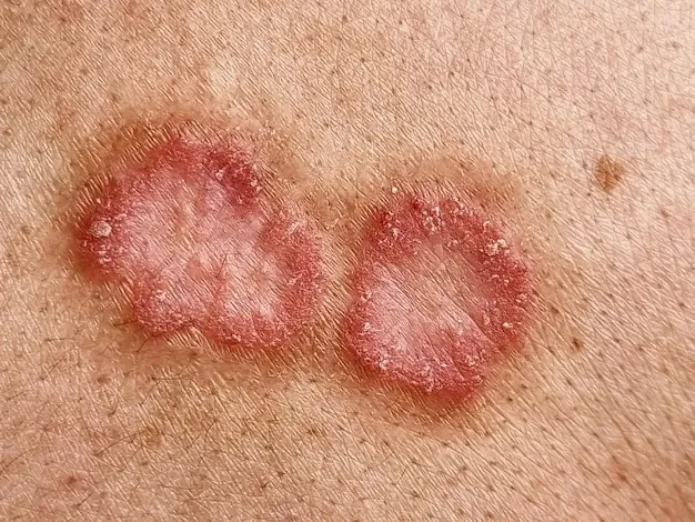
Fig. 4.6: Tinea corporis
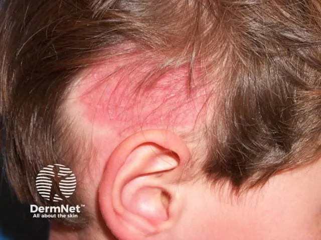
Fig. 4.7: Tinea capitis
Image sourced from DermNet.
-
Management:
-
Topical antifungals (clotrimazole, terbinafine) for skin.
-
Oral antifungals (griseofulvin, terbinafine) for scalp/nails.
-
Treat contacts and decontaminate shared items.
-
6. Candidiasis
Section titled “6. Candidiasis”-
Pathophysiology:
- Overgrowth of Candida albicans, especially in moist areas or the immunosuppressed.
-
Diagnosis:
-
Clinical: erythematous patches with satellite lesions.
-
KOH prep or culture.
-
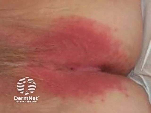
Fig. 4.8: Candidal vulvovaginitis
Image sourced from DermNet.
-
Management:
-
Topical nystatin or azoles (e.g., clotrimazole).
-
Oral fluconazole for systemic or recurrent cases.
-
Address predisposing factors (e.g., diabetes, antibiotics).
-
Parasitic Infection
Section titled “Parasitic Infection”7. Scabies
Section titled “7. Scabies”-
Pathophysiology:
-
Infestation by the Sarcoptes scabiei mite.
-
Hypersensitivity reaction to mite proteins.
-
-
Diagnosis:
-
Clinical: intense nocturnal pruritus; burrows in web spaces, axillae, and genitals.
-
Skin scraping with microscopy to confirm mites/eggs.
-
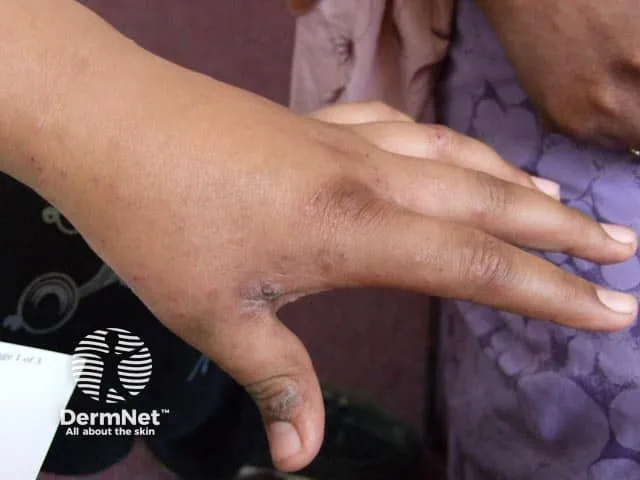
Fig. 4.9: Interdigital scaling in the first web space of the fingers
Image sourced from DermNet.
-
Management:
-
Topical permethrin 5% cream (apply overnight, repeat in 1 week).
-
Oral ivermectin for crusted or resistant cases.
-
Treat all close contacts simultaneously.
-
Wash bedding and clothing in hot water.
-
Essential Learning Notes
Section titled “Essential Learning Notes”Table 4.1: Essential Learning Notes — Skin Infections
| Category | Condition | Pathophysiology | Clinical Features | Diagnosis | Treatment |
|---|---|---|---|---|---|
| Bacterial | Impetigo | Superficial infection of the epidermis, commonly by Staphylococcus aureus or Streptococcus pyogenes | Honey-coloured crusts, often on face and limbs; non-bullous (more common) or bullous types | Clinical; Gram stain/culture if needed | Topical mupirocin (mild); oral antibiotics (e.g., cloxacillin, cephalexin) for extensive cases |
| Cellulitis | Infection of dermis and subcutaneous tissue, usually by Streptococcus pyogenes or S. aureus | Erythema, warmth, swelling, tenderness; may have fever, lymphangitis | Clinical; blood cultures if febrile/systemically ill | Oral/IV antibiotics (e.g., amoxicillin-clavulanate, clindamycin); elevation and rest | |
| Viral | Herpes Simplex | Reactivation of latent HSV-1 or HSV-2 in sensory ganglia | Grouped vesicles on erythematous base; painful; may recur | Tzanck smear (multinucleated giant cells), PCR, serology | Oral acyclovir, valacyclovir; topical antivirals for mild cases |
| Varicella (Chickenpox) | Primary infection with varicella-zoster virus (VZV) | Crops of vesicles on erythematous base (“dew drop on a rose petal”); pruritic; systemic symptoms | Clinical; PCR or serology if uncertain | Supportive care; acyclovir for high-risk or immunocompromised patients | |
| Fungal | Tinea (Dermatophytosis) | Infection by dermatophytes (Trichophyton, Microsporum, Epidermophyton) affecting keratinized tissue | Annular, scaly plaques with central clearing; varies by site (e.g., tinea corporis, pedis, capitis) | KOH prep, fungal culture | Topical antifungals (e.g., clotrimazole); oral terbinafine or griseofulvin for scalp/nail involvement |
| Candidiasis | Overgrowth of Candida albicans, especially in moist areas or immunocompromised hosts | Erythematous patches with satellite lesions; oral thrush, intertrigo, genital involvement | KOH prep, culture | Topical nystatin or azoles; oral fluconazole for systemic or recurrent cases | |
| Parasitic | Scabies | Infestation by Sarcoptes scabiei mite; hypersensitivity reaction to mite proteins | Intense pruritus (worse at night), burrows, papules in web spaces, wrists, axillae, genitals | Clinical; skin scraping with microscopy | Topical permethrin 5%; oral ivermectin for severe cases; treat close contacts and decontaminate linens |
Key Learning Points
Section titled “Key Learning Points”-
Recognize the pattern of lesion distribution and evolution (e.g., vesicles in HSV vs. crusts in impetigo).
-
Understand host factors (e.g., immunosuppression, hygiene, exposure) that influence infection risk.
-
Emphasize infection control: hand hygiene, contact precautions, and treatment of contacts (e.g., scabies).
-
Know when to escalate care: systemic symptoms, immunocompromised host, treatment failure.
Multiple Choice Questions
Section titled “Multiple Choice Questions”Tap an answer to check yourself — the correct option and an explanation appear once you choose.
Q1 A 6-year-old boy presents with honey-coloured crusted lesions around his mouth. He is otherwise well. What is the most appropriate first-line treatment?
The honey-coloured crusts indicate non-bullous impetigo. For localized, uncomplicated disease, topical mupirocin is first-line — as effective as oral antibiotics for limited impetigo, with fewer systemic side effects and less resistance. Oral flucloxacillin is reserved for extensive or complicated infection, acyclovir treats herpetic lesions (no honey crusts), and topical hydrocortisone can worsen a bacterial infection.
Q2 Which of the following features would most likely indicate a need for hospital admission in a patient with cellulitis?
Rapid progression with fever and hypotension signals systemic sepsis and warrants admission for IV antibiotics and monitoring. Mild, localized erythema and warmth without systemic features can usually be managed with oral antibiotics as an outpatient.
Q3 Which of the following is the most appropriate diagnostic test for confirming herpes simplex infection?
A Tzanck smear shows multinucleated giant cells characteristic of HSV (and VZV); PCR is more sensitive where available. KOH preparation is used for fungal hyphae, Gram stain for bacteria, and Wood’s lamp for certain fungal and pigmentary conditions.
Q4 A 22-year-old athlete presents with an annular, scaly lesion with central clearing on his thigh. What is the most likely diagnosis?
An annular, scaly plaque with central clearing is classic for tinea corporis (a dermatophyte infection); confirm with a KOH preparation. Psoriasis gives well-demarcated plaques with silvery scale, eczema is ill-defined and intensely itchy, and candidiasis favours moist flexures with satellite lesions.
Q5 Which of the following is the most appropriate management for scabies?
Topical permethrin 5% is first-line for scabies — applied overnight and repeated after one week, with simultaneous treatment of all close contacts and decontamination of bedding and clothing. Clotrimazole and fluconazole are antifungals, and acyclovir is an antiviral.
OSCE Stations
Section titled “OSCE Stations”OSCE 1: Impetigo Counselling
Section titled “OSCE 1: Impetigo Counselling”Scenario: You are a medical student in a GP clinic. A mother brings her 5-year-old child with crusted lesions around the nose. The GP has diagnosed impetigo.
Task: Counsel the mother on the nature of the condition, the treatment plan, infection control at home and school, and when to return for follow-up.
Self-assess against checklist
Tick each point you covered, then check your score.
- Introduces self and confirms patient/carer identity; uses clear, simple language appropriate for the parent.
- Explains that impetigo is a superficial bacterial infection of the outer layer of the skin (epidermis), most commonly caused by Staphylococcus aureus (which causes the bullous type) or Streptococcus pyogenes (which causes the more common non-bullous type).
- Describes the characteristic appearance: honey-coloured crusted lesions, most frequently around the nose and mouth; explains the two forms — non-bullous (crusted) and bullous (fluid-filled blisters).
- Explains how impetigo spreads: through direct contact with the lesions or via contaminated objects (fomites) such as towels, flannels, and clothing.
- Outlines the treatment plan: for mild, localized disease, topical mupirocin or fusidic acid applied directly to the lesions is first-line; for extensive involvement or systemic symptoms, oral antibiotics (e.g., cloxacillin or cephalexin) are required.
- Explains that a swab for culture should be taken if the infection is resistant to initial treatment or if it is recurrent, to guide antibiotic choice.
- Advises the child must stay away from school or daycare until the lesions are non-infectious — typically when crusts have dried and healed, or after 48 hours of antibiotic treatment.
- Advises strict handwashing for the child and all household members, especially before and after touching the lesions or applying treatment.
- Advises against sharing towels, flannels, clothing, or any items that come into contact with the lesions.
- Explains that siblings or close household contacts should be monitored for new lesions and assessed promptly if they develop similar symptoms.
- Advises to return if lesions are spreading, not improving after treatment, or if the child develops fever or systemic symptoms — which may indicate need for oral antibiotics.
OSCE 2: Scabies Diagnosis and Management
Section titled “OSCE 2: Scabies Diagnosis and Management”Scenario: A 19-year-old hostel student presents with intense itching, especially at night. Examination reveals burrows and papules in the web spaces of the fingers.
Task: Take a focused history, explain the diagnosis, and outline treatment and prevention for the patient and contacts.
Self-assess against checklist
Tick each point you covered, then check your score.
- Introduces self and confirms patient identity; reassures in a non-stigmatizing manner.
- Takes a focused history: onset and character of itch (specifically asking whether it is worse at night — nocturnal pruritus is the hallmark), distribution of lesions, duration of symptoms.
- Asks about close contacts with similar symptoms: roommates, hostel residents, household members, or sexual contacts.
- Examines (or describes examination findings): characteristic burrows and papules in the web spaces of the fingers, wrists, axillae, and genital area.
- Explains the diagnosis: scabies is an infestation by the Sarcoptes scabiei mite, which burrows into the skin; the intense itching is caused by a hypersensitivity reaction to mite proteins, eggs, and faeces.
- Explains that the itch is typically worse at night because mite activity increases with warmth under bedclothes.
- Explains how scabies spreads: through prolonged direct skin-to-skin contact; living in close quarters such as hostels increases transmission risk.
- Explains that a skin scraping with microscopy can confirm the diagnosis by identifying mites or eggs, if clinical diagnosis is uncertain.
- Explains the treatment: topical permethrin 5% cream is first-line — applied from the neck down to the entire body surface, left on overnight, and repeated after one week.
- Explains that oral ivermectin is used for crusted (Norwegian) scabies or cases resistant to topical treatment.
- Emphasises that all close contacts must be treated simultaneously, even if they are not yet symptomatic, to prevent re-infestation.
- Advises that all bedding, clothing, and towels used in the previous 72 hours must be washed in hot water and thoroughly dried.
- Warns that itching may persist for several weeks after successful treatment due to the ongoing hypersensitivity reaction, and that this does not necessarily mean treatment failure.
- Advises to return if symptoms persist beyond 2–4 weeks despite correct treatment, suggesting possible re-exposure or treatment failure.
Tutorial & Group-Work Prompts
Section titled “Tutorial & Group-Work Prompts”-
Case vignette: A 35-year-old diabetic woman presents with a red, warm, swollen leg and fever. Prompt: What is your differential diagnosis? What investigations and treatment would you initiate?
-
Visual diagnosis: Show images of tinea corporis, herpes simplex, and impetigo. Prompt: Identify each condition and justify your diagnosis based on lesion morphology.
-
Role play: One student plays a patient with varicella, another the doctor. Prompt: Practise explaining the diagnosis, home care, and when to seek medical attention.