Module 7: Drug Reactions and Emergencies
Learning Objectives
Section titled “Learning Objectives”By the end of this module, students should be able to:
-
Explain the immunological and non-immunological mechanisms underlying drug-induced skin reactions.
-
Identify clinical patterns of drug eruptions, SJS/TEN, urticaria, and anaphylaxis.
-
Differentiate between benign and life-threatening drug reactions based on clinical features.
-
Interpret relevant investigations to support diagnosis and monitor complications.
-
Initiate emergency management protocols for severe reactions, including anaphylaxis and SJS.
-
Educate patients on drug allergy documentation, avoidance strategies, and emergency preparedness (e.g., adrenaline auto-injector use).
1. Drug Eruptions
Section titled “1. Drug Eruptions”Pathophysiology:
-
Type I (IgE-mediated): Immediate hypersensitivity (e.g., urticaria, anaphylaxis).
-
Type IV (T-cell mediated): Delayed hypersensitivity (e.g., morbilliform rash, DRESS, SJS).
-
Non-immunologic: Direct mast cell degranulation (e.g., opioids, radiocontrast).
Diagnosis:
-
History: Drug exposure within 1–3 weeks; new or recently changed medications.
-
Clinical types:
-
Morbilliform eruption: Symmetrical maculopapular rash, trunk → limbs.
-
Fixed drug eruption: Recurrent, well-demarcated erythematous plaques.
-
DRESS: Fever, rash, eosinophilia, lymphadenopathy, hepatitis.
-
-
Investigations:
-
CBC (eosinophilia in DRESS), LFTs, renal function.
-
Skin biopsy if diagnosis is uncertain.
-
Management:
-
Stop the suspected drug immediately.
-
Mild: antihistamines, topical corticosteroids.
-
Severe (e.g., DRESS): systemic corticosteroids, hospital admission.
-
Document allergy in records and educate the patient.
Clinical Notes on Drug Eruptions
Section titled “Clinical Notes on Drug Eruptions”-
Fixed drug eruptions appear as oval erythematous patches that recur at the same site with re-exposure to the drug. Common sites include the glans penis, palms, soles, lips, neck, back, and face.
-
Bullous (blistering) fixed drug eruptions can occur, for example, due to sulphonamide antibiotics.
-
These lesions often heal with residual hyperpigmentation.
-
Drug eruption (drug rash or drug reaction) is a cutaneous adverse reaction to medications. It ranges from mild (red, itchy rash) to severe life-threatening reactions (e.g., Stevens-Johnson syndrome [SJS], toxic epidermal necrolysis [TEN]).
-
Approximately 2% of new prescriptions cause drug eruptions.
-
Common drugs causing eruptions include:
-
Antibiotics: penicillins, sulphonamides (sulfa drugs), tetracyclines
-
Anti-inflammatory drugs: NSAIDs like ibuprofen, naproxen
-
Anticonvulsants: phenytoin, carbamazepine
-
Chemotherapeutic agents
-
Psychotropic drugs
-
Hypertension drugs: diuretics, ACE inhibitors
-
Gout medication: allopurinol
-
HIV medications
-
Iodine-containing contrast media
-
-
Prompt identification and cessation of the offending drug is the main treatment.
-
Mild cases may be treated with emollients and oral antihistamines; severe reactions require specialist care.
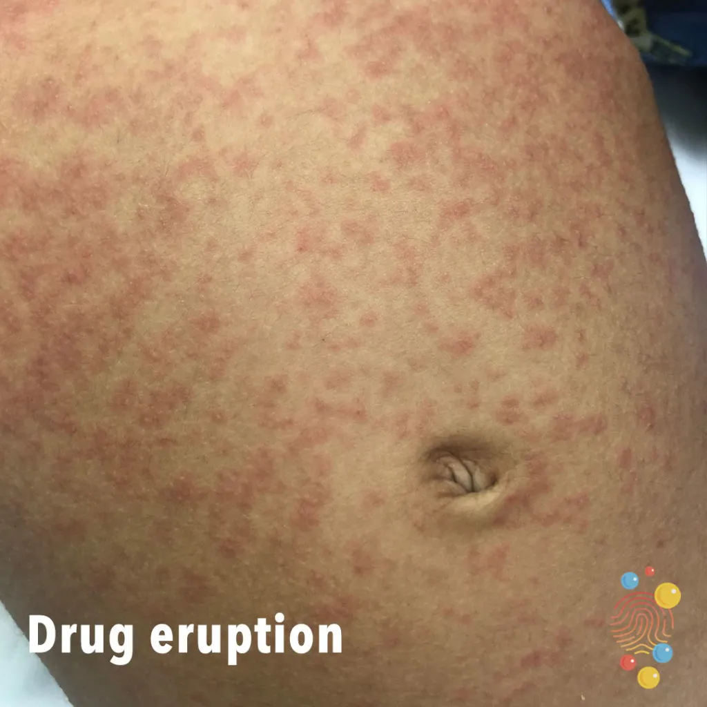
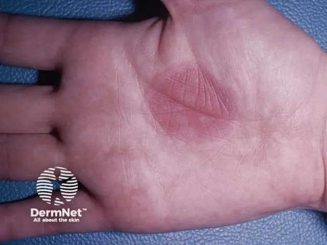
Fig. 7.1: Drug eruption
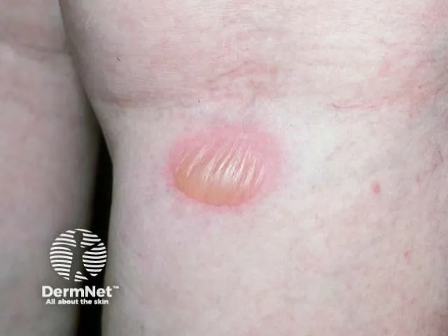
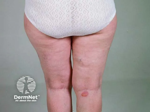
Fig. 7.2: Fixed drug eruption (due to sulphonamide)
Image sourced from DermNet.
2. Stevens-Johnson Syndrome (SJS)
Section titled “2. Stevens-Johnson Syndrome (SJS)”Pathophysiology:
-
Severe mucocutaneous reaction, usually drug induced.
-
Cytotoxic T-cell–mediated apoptosis of keratinocytes.
-
Common triggers: sulphonamides, allopurinol, carbamazepine, lamotrigine, NSAIDs.
Diagnosis:
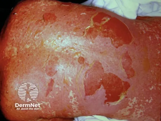
Fig. 7.3: Severe cutaneous reaction (SJS/TEN)
Image sourced from DermNet.
-
Prodrome: Fever, malaise, sore throat.
-
Skin: Targetoid lesions → blistering → epidermal detachment (<10% BSA in SJS).
-
Mucosal involvement: Oral, ocular, genital.
-
Investigations:
-
CBC, LFTs, renal function, electrolytes.
-
Skin biopsy: full-thickness epidermal necrosis.
-
Management:
-
Emergency admission to ICU/burns unit.
-
Immediate withdrawal of offending drug.
-
Supportive care:
-
Fluids, electrolytes, wound care, pain control.
-
Ophthalmology and urology consults.
-
-
Consider IVIG or cyclosporine in early stages (evidence evolving).
-
Avoid corticosteroids unless early and under specialist care.
Key Points: SJS Treatment
Section titled “Key Points: SJS Treatment”-
Immediate hospitalization is required, often in intensive care or burn units.
-
The first crucial step is to stop all potentially causative medications.
-
Supportive care includes:
-
Fluid replacement to manage dehydration from skin loss.
-
Nutritional support, sometimes via a nasogastric tube.
-
Wound care with gentle cleaning, use of petroleum jelly or medicated dressings.
-
Pain management using appropriate medications.
-
Eye care under the supervision of an ophthalmologist to prevent eye complications.
-
-
Medications that may be used:
-
Topical steroids to reduce inflammation.
-
Systemic corticosteroids and immunosuppressants like cyclosporine in some cases.
-
Intravenous immunoglobulin (IVIG) to modulate immune response.
-
Antibiotics only if secondary infections occur.
-
-
Monitoring and management of complications such as infections and toxic epidermal necrolysis is essential.
-
Recovery can take several days to months depending on severity.
3. Anaphylaxis and Urticaria
Section titled “3. Anaphylaxis and Urticaria”Pathophysiology:
Anaphylaxis:
- IgE-mediated Type I hypersensitivity → mast cell degranulation → systemic vasodilation, bronchospasm, shock.
Urticaria:
-
Histamine-mediated dermal oedema.
-
Acute (<6 weeks): often allergic.
-
Chronic (>6 weeks): autoimmune or idiopathic.
Diagnosis:
Anaphylaxis:
-
Rapid onset of:
-
Skin/mucosal symptoms (urticaria, angioedema)
-
Respiratory compromise (wheeze, stridor)
-
Hypotension or syncope
-
-
Common triggers: drugs, food, insect stings.
Urticaria:
-
Transient, itchy wheals ± angioedema.
-
No mucosal involvement unless part of anaphylaxis.
Management:
Anaphylaxis:
-
IM adrenaline 0.5 mg (1:1000) into lateral thigh immediately.
-
Airway support, high-flow oxygen, IV fluids.
-
Adjuncts: antihistamines, corticosteroids.
-
Observe for biphasic reaction (4–6 hours).
-
Prescribe adrenaline auto-injector and refer to the allergy clinic.
Urticaria:
-
Remove trigger if known.
-
Non-sedating antihistamines (cetirizine, loratadine).
-
Short course of oral corticosteroids if severe.
-
Chronic urticaria: consider autoimmune screen, H. pylori testing, referral.
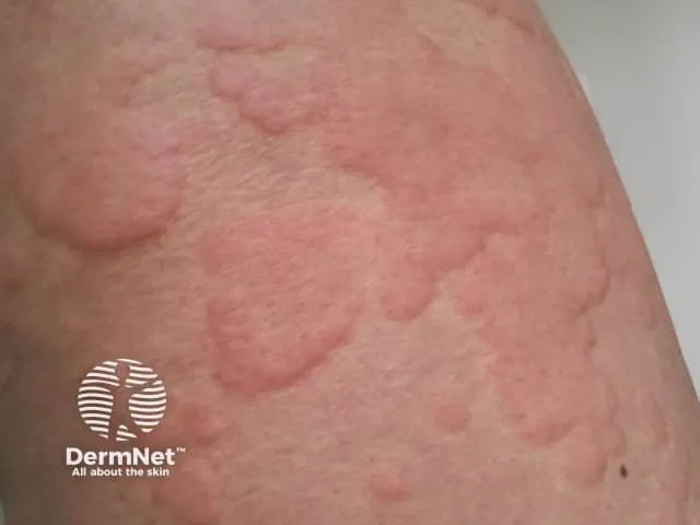
Fig. 7.4: Urticaria
Image sourced from DermNet.
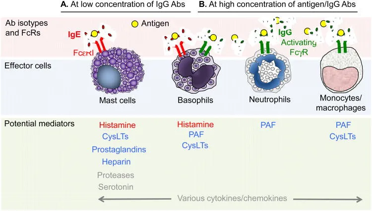
Fig. 7.5: Pathophysiology of anaphylaxis
Reber LL, Hernandez JD, Galli SJ. The pathophysiology of anaphylaxis. J Allergy Clin Immunol. 2017 Aug;140(2):335-348. doi: 10.1016/j.jaci.2017.06.003.
Multiple Choice Questions
Section titled “Multiple Choice Questions”Tap an answer to check yourself — the correct option and an explanation appear once you choose.
Q1 A 25-year-old man develops a widespread maculopapular rash 10 days after starting amoxicillin for a sore throat. He is afebrile and systemically well. What is the most likely diagnosis?
A symmetrical maculopapular rash appearing 7–14 days after starting an antibiotic in an otherwise well patient is the classic presentation of a morbilliform (exanthematous) drug eruption. Urticaria produces transient wheals, fixed drug eruption recurs at the same site, and SJS requires mucosal involvement and epidermal detachment with systemic illness.
Q2 Which of the following is the most appropriate initial step in managing a suspected drug eruption?
Discontinuing the offending drug is the first and most important step in managing any drug eruption. Systemic corticosteroids are reserved for severe reactions such as DRESS. Skin biopsy is a useful confirmatory investigation but not the immediate priority. Antibiotics are not indicated for a drug-induced rash.
Q3 A 40-year-old woman presents with fever, sore throat, and painful oral ulcers. She has dusky targetoid lesions on her trunk and arms. She started lamotrigine 2 weeks ago. What is the most likely diagnosis?
The combination of mucosal erosions (oral ulcers), targetoid skin lesions, fever, and a recent new anticonvulsant is the hallmark presentation of SJS. Erythema multiforme is generally less severe with no mucosal detachment at scale. Fixed drug eruption recurs at the same site and lacks mucosal involvement. DRESS is characterised by eosinophilia, lymphadenopathy, and organ involvement rather than targetoid blistering.
Q4 Which of the following is the most appropriate management for Stevens-Johnson Syndrome?
SJS is a dermatological emergency requiring immediate admission — ideally to an ICU or burns unit — with prompt withdrawal of the causative drug and intensive supportive care (fluid replacement, wound care, eye care). Oral antihistamines are inadequate. Antibiotics are only used if secondary infection develops. Continuing the drug risks progression to TEN and death.
Q5 A 35-year-old man develops sudden-onset wheezing, hypotension, and urticaria after receiving an IV antibiotic. What is the first-line treatment?
IM adrenaline (0.5 mg of 1:1000 solution) into the lateral thigh is the first-line treatment for anaphylaxis and must be given immediately. Corticosteroids and antihistamines are useful adjuncts but do not act quickly enough to reverse life-threatening bronchospasm and vascular collapse. IV fluids support blood pressure but do not address the underlying mediator release.
Q6 Which of the following is true regarding chronic urticaria?
Chronic urticaria (lasting more than 6 weeks) is most commonly autoimmune or idiopathic rather than IgE-mediated. It does not always require systemic steroids — first-line therapy is non-sedating antihistamines. IgE-mediated urticaria is typical of acute episodes (under 6 weeks).
Short Answer Questions
Section titled “Short Answer Questions”SAQ 1: Drug Eruption
Section titled “SAQ 1: Drug Eruption”SAQ
List four clinical types of drug eruptions and provide one example of a drug commonly associated with each.
Reveal model answer
-
Morbilliform eruption — e.g., amoxicillin
-
Fixed drug eruption — e.g., NSAIDs
-
DRESS syndrome — e.g., carbamazepine
-
Urticaria/angioedema — e.g., penicillin
SAQ 2: Anaphylaxis
Section titled “SAQ 2: Anaphylaxis”SAQ
Outline the immediate management steps for a patient presenting with anaphylaxis.
Reveal model answer
-
Administer IM adrenaline 0.5 mg into the lateral thigh immediately.
-
Provide high-flow oxygen.
-
Establish IV access and give IV fluids.
-
Administer antihistamines and corticosteroids as adjuncts.
-
Monitor for biphasic reaction.
-
Prescribe adrenaline auto-injector and refer to the allergy clinic.
OSCE Stations
Section titled “OSCE Stations”OSCE 1: Drug Eruption Counselling
Section titled “OSCE 1: Drug Eruption Counselling”Scenario: A 22-year-old woman presents with a widespread itchy rash after starting trimethoprim-sulfamethoxazole for a UTI. She is systemically well.
Task: Take a focused drug history, explain the likely diagnosis, and advise on management and future precautions.
Self-assess against checklist
Tick each point you covered, then check your score.
- Identifies timing of drug exposure relative to rash onset.
- Explains morbilliform drug eruption in plain terms.
- Advises stopping the causative drug.
- Recommends antihistamines and topical steroids for symptom relief.
- Emphasises allergy documentation in medical records and avoidance of the drug class in future.
OSCE 2: SJS Emergency Management
Section titled “OSCE 2: SJS Emergency Management”Scenario: A 45-year-old man presents with fever, mucosal erosions, and a blistering rash after starting allopurinol. You are a medical student in the ED.
Task: Recognise the emergency, outline immediate management steps, and communicate with the patient and team.
Self-assess against checklist
Tick each point you covered, then check your score.
- Recognises Stevens-Johnson Syndrome and immediately stops the causative drug.
- Calls for urgent admission to ICU or burns unit.
- Orders relevant investigations: CBC, LFTs, renal function, electrolytes, skin biopsy.
- Outlines supportive care plan: IV fluids, wound care, pain management, eye care.
- Communicates seriousness to the patient and advocates for multidisciplinary involvement (ophthalmology, urology).
OSCE 3: Anaphylaxis Response
Section titled “OSCE 3: Anaphylaxis Response”Scenario: A patient develops sudden urticaria, wheeze, and hypotension after a penicillin injection in the clinic.
Task: Recognise and manage the emergency, administer appropriate treatment, and communicate with the team and patient.
Self-assess against checklist
Tick each point you covered, then check your score.
- Recognises anaphylaxis based on urticaria, bronchospasm, and hypotension.
- Administers IM adrenaline promptly into the lateral thigh.
- Calls for help and monitors vital signs.
- Starts high-flow oxygen and IV fluids.
- Documents the reaction and advises the patient on future avoidance and adrenaline auto-injector use.