Module 8: Paediatric and Geriatric Skin Disorders
Learning Objectives
Section titled “Learning Objectives”By the end of this module, students should be able to:
General Objectives
-
Describe the anatomical and physiological differences in paediatric and geriatric skin.
-
Recognize common dermatological conditions specific to children and older adults.
-
Understand the pathophysiology underlying age-specific skin disorders.
-
Differentiate between normal age-related changes and pathological conditions.
-
Formulate appropriate diagnostic and management plans tailored to age and comorbidities.
-
Communicate effectively with caregivers and elderly patients regarding skin care and treatment adherence.
Section A: Paediatric Dermatology
Section titled “Section A: Paediatric Dermatology”Skin Characteristics in Children
Section titled “Skin Characteristics in Children”-
Thinner epidermis and stratum corneum → increased permeability.
-
Immature immune system → higher susceptibility to infections.
-
Higher surface area-to-body weight ratio → increased risk of systemic absorption of topical agents.
1. Atopic Dermatitis (Eczema)
Section titled “1. Atopic Dermatitis (Eczema)”Dupilumab is now recommended for moderate-to-severe cases unresponsive to topical therapy.
Pathophysiology:
-
Genetic predisposition (e.g., filaggrin mutation) → impaired skin barrier.
-
Th2-dominant immune response → chronic inflammation.
-
Environmental triggers: allergens, irritants, infections.
Diagnosis:
-
Chronic, relapsing pruritic rash.
-
Infants: cheeks, scalp, extensor surfaces.
-
Children: flexural areas (elbows, knees).
-
Associated with asthma and allergic rhinitis.
Management:
-
Emollients: cornerstone of therapy.
-
Topical corticosteroids: mild to moderate potency.
-
Antihistamines: for sleep-disturbing pruritus.
-
Education: trigger avoidance, bathing practices, long-term care.
2. Diaper Dermatitis
Section titled “2. Diaper Dermatitis”Pathophysiology:
-
Irritant contact dermatitis from prolonged exposure to urine/faeces.
-
Secondary Candida albicans infection is common.
Diagnosis:
-
Erythema, scaling in the diaper area.
-
Candida: satellite lesions, involvement of skin folds.
Management:
-
Frequent diaper changes.
-
Barrier creams (zinc oxide).
-
Topical antifungals (e.g., nystatin) if Candida suspected.
-
Avoid irritant wipes.
3. Tinea Capitis
Section titled “3. Tinea Capitis”Pathophysiology:
-
Dermatophyte infection of scalp and hair shafts.
-
Common organisms: Trichophyton tonsurans, Microsporum canis.
Diagnosis:
-
Scaly patches with alopecia.
-
“Black dot” or inflammatory kerion.
-
KOH prep, fungal culture.
Management:
-
Oral antifungals: griseofulvin or terbinafine.
-
Topical antifungals: adjunctive only.
-
Treat contacts and decontaminate combs/hats.
4. Impetigo
Section titled “4. Impetigo”Pathophysiology:
-
Superficial bacterial infection by S. aureus or S. pyogenes.
-
Spread via direct contact or fomites.
Diagnosis:
-
Honey-coloured crusts, often on the face.
-
Bullous or non-bullous types.
Management:
-
Topical mupirocin for localized lesions.
-
Oral antibiotics (e.g., cloxacillin, cephalexin) for extensive disease.
-
Hygiene education to prevent spread.
Section B: Geriatric Dermatology
Section titled “Section B: Geriatric Dermatology”Skin Characteristics in the Elderly
Section titled “Skin Characteristics in the Elderly”-
Thinning of epidermis and dermis.
-
Decreased collagen, elastin, and sebaceous gland activity.
-
Reduced immune surveillance and delayed wound healing.
-
Increased risk of skin tears, infections, and malignancies.
1. Xerosis (Dry Skin)
Section titled “1. Xerosis (Dry Skin)”Pathophysiology:
-
Reduced lipid content and sweat gland activity.
-
Exacerbated by cold weather, frequent bathing, and harsh soaps.
Diagnosis:
-
Dry, flaky, itchy skin, especially on lower legs.
-
May lead to eczema craquelé or secondary infection.
Management:
-
Emollients (urea-based or petrolatum).
-
Avoid hot showers and harsh soaps.
-
Humidifiers in dry environments.
2. Senile Purpura
Section titled “2. Senile Purpura”Pathophysiology:
-
Fragility of dermal blood vessels due to collagen loss.
-
Minor trauma causes extravasation of blood.
Diagnosis:
-
Non-palpable purpura on forearms and hands.
-
No systemic symptoms.
Management:
-
Reassurance (benign).
-
Protective clothing.
-
Avoid anticoagulants/steroids if not essential.
3. Seborrhoeic Keratosis
Section titled “3. Seborrhoeic Keratosis”Pathophysiology:
-
Benign proliferation of immature keratinocytes.
-
Common with aging; not premalignant.
Diagnosis:
-
Waxy, “stuck-on” appearance.
-
Pigmented or skin-coloured plaques.
Management:
-
No treatment unless symptomatic or cosmetic.
-
Cryotherapy or curettage if removal desired.
4. Skin Malignancies
Section titled “4. Skin Malignancies”Pathophysiology:
-
Cumulative UV exposure → DNA damage.
-
Immunosenescence increases risk.
Diagnosis:
-
BCC: pearly papule with telangiectasia.
-
SCC: scaly, crusted lesion; may ulcerate.
-
Melanoma: ABCDE criteria.
Management:
-
Biopsy for diagnosis.
-
Surgical excision is the mainstay.
-
Sun protection and regular skin checks.
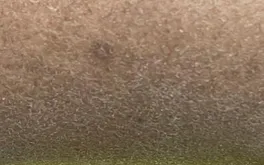
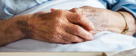
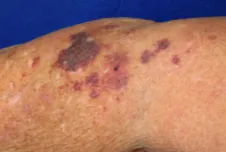
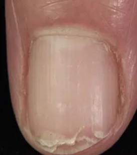
Fig. 8.1: Common Geriatric Skin Disorders
- Xerosis (dry skin) with scaling
- Bruising
- Senile purpura
- Brittle nail
- Onychomycosis
Multiple Choice Questions
Section titled “Multiple Choice Questions”Tap an answer to check yourself — the correct option and an explanation appear once you choose.
Q1 A 2-year-old child presents with itchy, dry, red patches on the cheeks and extensor surfaces. There is a family history of asthma. What is the most likely diagnosis?
Atopic dermatitis (eczema) classically presents in infancy with pruritic erythematous patches on the cheeks and extensor surfaces, often with a family history of atopy (asthma, allergic rhinitis). Seborrhoeic dermatitis produces greasy yellow scales in sebaceous-rich areas, psoriasis gives well-demarcated silvery plaques, and tinea corporis shows annular scaly lesions.
Q2 A 7-year-old boy presents with a scaly patch of hair loss on the scalp. There are black dots and mild inflammation. What is the most appropriate treatment?
Tinea capitis requires systemic antifungal therapy because topical agents do not penetrate the hair shaft adequately. Oral griseofulvin is the traditional first-line agent in children; terbinafine is an alternative. Topical antifungals alone are insufficient and are used only as adjuncts. Fluconazole is not a standard first-line choice for tinea capitis.
Q3 An 80-year-old woman presents with painless purple patches on her forearms. She is on aspirin and warfarin. What is the most likely diagnosis?
Senile purpura is caused by age-related loss of perivascular collagen support, leading to non-palpable purpuric patches on sun-exposed areas such as the forearms and dorsum of the hands after minor trauma. It is a benign, diagnosis of exclusion. Vasculitis is typically palpable, traumatic ecchymosis is usually a single larger lesion with a clear history, and thrombocytopenia would show widespread petechiae with a low platelet count.
Q4 Which of the following features is most suggestive of malignant melanoma?
Malignant melanoma is identified using the ABCDE criteria: Asymmetry, Border irregularity, Colour variation, Diameter >6 mm, and Evolution (change over time). A rapidly changing lesion with irregular borders is a hallmark red flag. A symmetrical uniform brown lesion is more consistent with a naevus, a pearly papule with telangiectasia describes basal cell carcinoma, and a rough scaly patch on sun-exposed skin suggests actinic keratosis or squamous cell carcinoma.
Short Answer Questions (SAQs)
Section titled “Short Answer Questions (SAQs)”SAQ 1: Paediatric Skin — Atopic Dermatitis
Section titled “SAQ 1: Paediatric Skin — Atopic Dermatitis”SAQ
List four key components in the management of atopic dermatitis in children.
Reveal model answer
-
Regular use of emollients to restore and maintain the skin barrier.
-
Topical corticosteroids for flare control (mild to moderate potency).
-
Avoidance of triggers (e.g., soaps, allergens, heat, synthetic fabrics).
-
Parental education on the chronic, relapsing nature of the condition and appropriate skin care routines.
SAQ 2: Geriatric Skin — Age-Related Changes
Section titled “SAQ 2: Geriatric Skin — Age-Related Changes”SAQ
Describe three physiological changes in aging skin and their clinical implications.
Reveal model answer
-
Thinning of epidermis and dermis → increased skin fragility and risk of skin tears with minor trauma.
-
Reduced sebaceous gland activity → decreased surface lipids leading to dry skin (xerosis) and pruritus.
-
Decreased immune surveillance → reduced immunosenescence increases susceptibility to infections and skin malignancies.
OSCE Case Scenarios
Section titled “OSCE Case Scenarios”OSCE 1: Paediatric Case — Diaper Dermatitis
Section titled “OSCE 1: Paediatric Case — Diaper Dermatitis”Scenario: A 9-month-old infant presents with a red rash in the diaper area. The rash involves the skin folds and has satellite lesions.
Task: Take a focused history and explain the diagnosis and management to the parent.
Self-assess against checklist
Tick each point you covered, then check your score.
- Identifies prolonged diaper use and recent diarrhoea as contributing factors.
- Recognises involvement of skin folds and satellite lesions as features of Candida superinfection.
- Explains diagnosis: Candida-associated diaper dermatitis.
- Recommends frequent diaper changes and application of barrier cream (zinc oxide).
- Prescribes topical antifungal (e.g., nystatin cream).
- Advises on hygiene practices and avoidance of irritant wipes.
- Provides safety-netting and advice on when to return for follow-up.
OSCE 2: Geriatric Case — Skin Tear and Xerosis
Section titled “OSCE 2: Geriatric Case — Skin Tear and Xerosis”Scenario: An 82-year-old nursing home resident presents with a skin tear on the forearm and complains of dry, itchy skin.
Task: Assess the skin and identify contributing factors. Explain preventive strategies to the caregiver.
Self-assess against checklist
Tick each point you covered, then check your score.
- Identifies xerosis and fragile aging skin as the underlying cause.
- Explains age-related physiological changes (collagen loss, thinning dermis) and their implications.
- Recommends regular application of emollients (urea-based or petrolatum).
- Advises on gentle handling, avoiding tight grips and friction.
- Recommends protective clothing (long sleeves) to reduce trauma risk.
- Addresses fall prevention and environmental safety in the care setting.
- Documents the skin tear and initiates appropriate wound care.
OSCE 3: Geriatric Case — Suspicious Pigmented Lesion
Section titled “OSCE 3: Geriatric Case — Suspicious Pigmented Lesion”Scenario: A 70-year-old man presents with a pigmented lesion on his back that has changed in size and colour over 3 months.
Task: Take a focused history and examine the lesion. Explain the likely diagnosis and next steps to the patient.
Self-assess against checklist
Tick each point you covered, then check your score.
- Takes a focused history including duration, rate of change, bleeding, and prior sun exposure.
- Applies ABCDE criteria: Asymmetry, Border irregularity, Colour variation, Diameter, Evolution.
- Raises clinical suspicion of malignant melanoma based on findings.
- Recommends urgent dermatology referral and excision biopsy for histological confirmation.
- Educates the patient on sun protection (broad-spectrum sunscreen, protective clothing, avoiding peak UV hours).
- Advises on skin self-examination and importance of early detection.
- Documents findings clearly and arranges timely follow-up.